

 PDF
PDF ePub
ePub Citation
 Print
Print


INTRODUCTION
The most common cause of foreign body diseases is coins in children. However, in adults, this can vary in different localities and there are some differences among different nations. In Western countries, impacted meat was prevalent (1) and this could be associated with esophageal structural abnormalities. In Asian countries, fish bones were the most prevalent cause (2) and this seemed related to Asian's eating habits.
An esophageal fish bone foreign body (FBFB) is common among the people of Jeju Island because the local people enjoy eating fish. Among the fish, they especially like to eat the fish named locally as "Jari" or "Jaridom", the damselfish or coralfish Chromis notata, which is a less than a 15 cm sized small fish. It inhabits the inshore and offshore coral or rocky reefs from 2 to 15 m depth and this fish is distributed in the northwest Pacific, including southern Japan, Taiwan, China, and southern Korea, including Jeju Island (3).
During our medical practice, we found that the FBFBs of damselfish are common and caused by exclusively a specific part of the fish's skeleton. Therefore, we conducted this study to determine which the parts of the fish skeleton was involved and to evaluate the clinical characteristics of patients with a FBFB from damselfish.
MATERIALS AND METHODS
Study group
We reviewed retrospectively the medical records, endoscopic photographs and radiologic findings of the patients who had endoscopic or surgical treatment for esophageal foreign body to find the rate of damselfish FBFB from March 2004 to March 2011 in Jeju National University Hospital. We collected the data on age and gender of the patients, the kinds of foreign body and the types of fish, the sizes and nature of the foreign bodies, the involved levels, the causative fish, the cooking recipes, evidence of mural penetration, the time delay from symptom onset to a hospital, and the admission durations.
Study protocol
The time delays between symptom onset and the hospital arrival were divided four groups as follows; within 24 hr, 48 hr, 72 hr, and over 72 hr. The types of fish bone were classified to four groups; linear bone, anal fin spine (AFS), skull bone and back bone according to the photographically assessed shapes. The skull bones included flat bones, jaw bones and gills. All the sharp and linear pin-like bones are grouped to the linear bones except AFS. The recipes were soup, hard-boiled, baked, sliced raw fish with sauce Jari mulhoe, salt-pickled, and steamed. Various Korean recipes that had a broth component were all classified to soup. The lodged sites were classified to four groups as 1) pharynx, 2) upper esophagus; the upper esophageal sphincter to 23 cm from the upper incisor, 3) mid esophagus; the aortic arch eminence to 31 cm from the upper incisor, and 4) lower esophagus; below the mid esophagus.
The simple X-rays and the computed tomography (CT) images were read by a radiologist. The CT findings were categorized to five groups; 1) those with negative finding, 2) visible fish bone without adjacent wall edema, 3) visible fish bone with adjacent wall edema, 4) visible mediastinal free air and 5) abscess and prominent mediastinitis.
Endoscopic evidence of pharyngeal and esophageal wall penetration was also evaluated. In the cases of surgical treatment without endoscopy, it was judged by the surgical records.
The monthly and yearly distributions of FBFB were also checked. Above procedures including reading the endoscopic photography and grouping the fish bones into different categories and judging the penetration were done by one person to reduce the interpersonal variation and to achieve consistency.
Statistical analyses
The statistical analyses were done using the SPSS for Windows (V. 13.0, SPSS, Chicago, IL, USA), and P values < 0.05 were considered to be significant. Descriptive statistics were used to summarize the characteristics of the cases and controls. Bivariate analysis was performed with chi-squared tests for the categorical variables and t-tests were used for the continuous variables as appropriate.
RESULTS
The types of foreign bodies and fishes
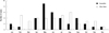
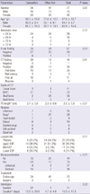
Our study included 126 cases of upper gastrointestinal foreign bodies: fish bones were most common and they were recorded in 77 cases 61.1%, meat in 26 cases 20.6%, other food material in 6 cases, chicken bones in 5 cases, drug in 4 cases; pills in 2, a press-through package in 2 and others in 8; nasogastric tube in 3, denture 2, tooth brush, sea shell and safety pin in one case each. The most common FBFB was damselfish and it accounted for 36 cases (46.8%). Others were armorclad rockfish in 14, houttuyn in 8, cutlassfish in 3, yellow porgy in 3, Alaska pollack in 2, horse mackerel in 2, unknown 4 and the others in 5 cases; shotted halibut, sawedged perch, rudder fish, black rockfish and redlip croaker in one case each (Table 1). The FBFB cases were common from April to August and they were most common in May, especially in damselfish FBFB case (Fig. 1).
Clinical characteristics of fish bone foreign body diseases
The gender distribution was 35 men and 42 women in all the FBFB disease cases, and 16 men and 20 women for the cases of damselfish FBFB disease (Table 2). The mean age (mean ± standard deviation) was 57.8 ± 12.7 yr. The men were younger than the women 54.1 ± 8.7 for men and 60.9 ± 14.7 for women.
The time delays to hospital arrival were grouped as within 24 hr (n = 57), 48 hr (n = 10), 72 hr (n = 5), and over 72 hr (n = 4). Except for 1 case each in the latter two groups, the remaining 8 cases were damselfish FBFB. The longest time delay to hospital arrival were 5 days in two cases.
Simple X-rays were taken in 67 cases and 7 cases had overt evidence of FBFB disease sensitivity: 10.5%. Of these 7 cases, 6 cases were damselfish FBFB disease (P = 0.04). CTs were taken in 44 cases and 43 cases had positive findings (sensitivity 97.7%). The positive groups were classified to four subgroups: only a FBFB was detected in 25 cases, FB induced tissue edema was detected in 3 cases, positive free gas was detected in 11 cases, and abscess or mediastinitis was detected in 4 cases. Of the 11 cases with positive free gas findings, 10 cases were damselfish FBFB and 3 out of the 4 cases of the abscess or mediastinitis group were damselfish FBFB. Damselfish FBFB diseases had more serious findings on CT (P = 0.04).
The shapes of the FBFBs were categorized to four groups: 11 linear bones, 33 anal fin spine-pterygiophore complexes (ASPC), 29 skull bones and 4 spinal bones. Except for one case of a skull bone FB, 28 cases were from fish other than damselfish. Of the total 36 cases of damselfish FBFB, the ASPC was involved in 32 cases (88.9%) and it was the most common. The mean length of the FBFBs was 2.51 ± 0.81 cm; damselfish 2.73 ± 0.77 cm, other fish 2.33 ± 0.81 cm.
In the damselfish FBFB group, one recipe was soup, 7 were hard-boiled, 6 were baked, 5 were sliced raw fish with sauce, 8 were salt-pickled and 2 were steamed. Sliced raw fish with sauce and salt-pickled are relatively unique recipes for damselfish. In the other FBFB group, soup (n = 27) was most common recipe and the 2 hard-boiled and 4 baked. The most common recipes were soup in the total FBFB cases. There were remarkable differences in the recipes between damselfish and the other FBFB groups (P < 0.01).
The lodged sites for the total FBFBs were the pharynx in 22 cases (28.6%), the upper esophagus in 35 cases (45.5%), the mid-esophagus in 11 cases (14.3%) and the lower esophagus in 9 cases (11.7%). The most common site was the upper esophagus. There was no statistical significance in the foreign body lodged site between the damselfish and other FBFB groups (P = 0.11).
In 33 cases (damselfish 23, other fishes 10), endoscopic evidence of mural penetrations by the FBFB was noticed. Because the damselfish FBFBs were exclusively ASPC that had sharp points on both ends, both sides of pharyngoesophageal penetration were seen in 14 cases of damselfish FBFBs, compared to 2 cases of other FBFBs (P < 0.01).
Among the 77 cases of FBFB disease, 75 cases were treated by endoscopy and the remaining two cases were surgically operated on. All the two operated cases were caused by a damselfish FBFB. Thirty one cases (40.3%) were hospitalized after FBFB removal and the mean number of days of hospital treatment was 10.3 ± 17.3 days. Hospital admission was more common in the damselfish FBFB group (n = 22) compared with the other FBFB group (n = 9) and the mean number of days of hospital treatment was longer in the damselfish FBFB group compared with that of the other FBFB group (12.6 ± 20.0 cm vs 4.7 ± 4.8 cm, respectively, P = 0.02).
Morphological characteristics of Chromis notata fish bone
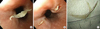
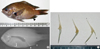
The scientific name of damselfish is Chromis notata. Among the many bony portions of damselfish causing esophageal fish bone foreign body, a uniquely-shaped linear bone portion was frequently noticed. Endoscopically, it was boomerang shaped and it had two sharp ends and a joint on the mid portion. It can penetrate both sides of the luminal wall of the esophagus and perforate it (Fig. 2). We examinated 18 damselfish that were caught in October to identify the specific boomerang-shaped, sharp portion of the damselfish bone. After boiling the damselfish, we took the meat off the bones. The typical shaped bone portion was found at the anal fin portion just posterior to the anus. One damselfish anal fin had 12 AFSs and the first two AFSs were ossified and very sharp. The first AFS was short, but the second AFS was very long and sharp. The first two AFSs were jointed to the inner pterygiophore. The first anal pterygiophore was also strongly ossified and long. Three portions of the bone were tightly adhered with joints. The mean length of the processed ASPC was 3.67 cm. They were not dislocated even after being hard-boiled, baked or roasted. They jointed with a wide angle and they flexed to 120°. The first and second AFSs were independently mobile. The tip of the pterygiophore was sharp. However, the AFSs were very sharp, hard and comparable to sewing needles (Fig. 3). Contrary to the very strong first and second AFSs, the pectoral and pelvic fins were relatively soft, and the first dorsal spines were short and easily dissociated from the pterygiophores.
The bone unit of the ASPC was very strong, dense and long enough to perforate both esophageal walls. The ASPC is linked by means of a simple joint and ligament. The ASPC is typically sharp at both ends. The spine side is sharper and harder. The long spine is the second AFS and the rudimentary small first AFS is also attached to it (Fig. 3). If the complex deeply penetrates the esophageal wall by the pterygiophore side, the FB removal becomes very difficult because the first AFS acts as an anchor in the esophageal wall during the foreign body removal.
DISCUSSION
In present study, fish bone was the most prevalent foreign body (61.1%) and meat was ranked second (20.6%); and this ranking was not different compared to other Asian country (2). Damselfish was the most prevalent source of FBFB. The high prevalence of damselfish FBFB may be due to 1) abundant and cheap, 2) traditionally favored fish, and 3) unique shape of the FBFB. In this study, it was unknown why the female patients were older than the male patients 60.9 ± 14.6 yr vs 54.1 ± 8.7 yr, respectively, and the trend was same in the damselfish and other fish FBFB disease groups.
Delayed hospital arrival is one of the poor prognostic factors. There was no significant difference of age and gender between early and later arrival groups. However, of 9 cases who were delayed over 48 hr, 8 cases (88.9%) were the patients of damselfish FBFB. We thought it was caused by the morphological characteristics of the damselfish FBFB; the both side penetrable shape of the FBFB, which enable decreased luminal pressure despite of both side perforation.
The sensitivity of plain radiography is low to detect foreign bodies, and early detection is depend on the bony calcification or kind of fishes (4, 5). CT is superior to plain radiographs for localizing and identifying foreign bodies and it is highly reliable for localizing foreign bodies in the esophagus (6). Some authors reported that the sensitivity and specificity of CT reached 78% and 96% respectively, and the positive and negative predictive value reached upto 75% and 97% respectively (7). In the present study, plain radiography detected only 7 fish bones in 67 cases (11.7%). A 6 of the 7 cases were caused by damselfish bones. The FBFBs detected by CT in 43 of 44 cases (97.7%). The missing case was caused by a 1-cm sized transparent small flat bone.
The local people usually eat raw flesh of medium sized fish as sashimi at first, and then boil the remaining fish head and back bones for soup for the medium sized fish. That is the reason why the flat fish skull bones were common in the other FBFBs. Exceptionally houttuyn is initially boiled for soup without eat sashimi. On the other hand, the majority of the local people cooks and eats damselfish as unboned state. Because the skull and back spine of damselfish are small and weak, the fishbone could be enough to chew and swallow without luminal lodgement. However the ASPC is too hard to be chewed. Of the total 36 cases of damselfish FBFB disease, 31 cases were caused by the ASPC. It is important that it has two sharp, bony ends that are long enough to penetrate deeply the esophageal wall.
Generally, esophageal perforation due to foreign body ingestion is rare and accounts for 1%-4% of the total reported cases (8). In one study, the pharyngeal location, fish bone, and radiolucency were associated with an increased incidence of complications in patients over 10 yr of age (9). The fish bones have a sharp polygonal or pin-like pointed structure and they can perforate or tear the esophageal wall. It can cause pneumomediastinum, mediastinitis, pneumothorax (10), tracheoesophageal fistula (11), major vascular damage (8, 12), and so far as to systemic air embolism in rare case (13). Upper esophageal and oropharyngeal FBFB can cause soft tissue infection and other complications including fistula formation and carotid arterial injury (14, 15). We already had several cases of pneumomediastinum, mediastinitis, and aortic injury.
Thoracic esophageal perforations carry a greater risk of morbidity and mortality than cervical esophageal perforations due to the operative risks (16). The overall mortality rate in the case of esophageal perforation is 22%, but the survival rate is up to 80%-90% when the primary repair is completed within 24 hr of rupture (17). Successful treatment of esophageal perforation is depends on the size of the perforation, the time delay to diagnosis and the underlying medical condition (18). In present study, only two cases underwent surgical operation because of abscess formation. In those cases, the time delay was 3 and 5 days before hospital arrival and the foreign body lodged to upper esophageal junction and hypopharynx. We had no case of mortality associated with FBFB disease and surgery. Except for the two cases, most of the cases successfully treated with conservative management.
Most of the small foreign bodies are naturally eliminated via the esophagus and gastrointestinal tract without symptom or complication (19). Therefore, nonoperative intervention is needed in 10%-20% of the cases; and surgical treatment is needed in 1% (20). The indications for surgery are peritoneal perforation, pneumothorax, clinical signs of systemic sepsis, respiratory insufficiency, shock and widespread of contamination due to a large perforation (21-24). In the other hand, the criteria for nonoperative treatment include early diagnosis, limited air leakage, not associated with visceral obstruction or malignancy, no evidence of septicemia (25-27), and the most reliable setting is locally contained and limited inflammatory changes surrounding the perforation (28). Antibiotics that active on both aerobes and anaerobes should be started because of the polymicrobic bacterial natures of the majority of cases and anaerobes predominance (29). In this study, many cases of FBFB due to the damselfish ASPC penetrated the esophageal wall. The majority of the perforations sealed with the FBFB itself and the perforation size were as small as a pin hole. Therefore, if the FBFB disease was diagnosed early and there were no signs and symptoms of mediastinitis, then medically supportive care would be sufficient.
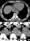
The damselfish have an anal fin with two spines and adhered to the fish body by the pterygiophore (3). The pterygiophore is ossified linear bone serves as roots of the anal fins, and muscles for the movement of the fins are attached (30). The esophagus runs with the major vessels in the thoracic cavity, and it has three physiologic narrowed points. The portion near the aortic arch is one of them. In this study, the aortic arch level was included in the mid esophagus and 14.3% of all the FBFBs were lodged in the site. Damselfish FBFBs were relatively common in the mid esophagus (22.2%). The damselfish FBFBs are sharp and long enough to perforate the aorta or other thoracic organs. Several cases involved in the aorta or major vessels (Fig. 4).
In conclusion, damselfish FBFBs are most common in the people of Jeju Island. The majority of the FBFBs are caused by the ASPC and this caused a more significant clinical course. It is important to be aware that a damselfish FBFB can damage major vessels. This study is the first report that a specific part of fish bone, the ASPC, is the major cause of FBFB disease and it can be dangerous. We recommend removing the ASPC during cleaning the damselfish befor e cooking it. An early diagnosis and treatment is important because a delayed diagnosis can cause severe complications and increase the need for surgery.
 XML Download