

 PDF
PDF ePub
ePub Citation
 Print
Print


INTRODUCTION
Reactive oxygen species (ROS) are well-known mediators of intracellular signaling cascades, and include free radicals, such as superoxide (O2-), hydroxyl radical and compounds such as hydrogen peroxide (H2O2) (1, 2). Because of the excessive ROS generation is very toxic to cell, cells also have intrinsic antioxidant systems that limit ROS action (2). Oxidative stress occurs when excessive ROS are generated that cannot be countered by intrinsic antioxidant system (2). Excessive ROS generation triggers diverse cellular dysfunctions, including lipid peroxidation, DNA mutagenesis, and cellular apoptosis or necrosis (2). ROS act as the key mediators for pathogenesis of various cardiovascular diseases with cardiomyocyte (CMC) hypertrophy, matrix remodeling, promotion of transcriptional factors, and cellular signal transduction abnormality (1, 3).
Thioredoxin-1 (Trx-1) is a ubiquitous, small molecular weight (12 kDa) protein, which was originally identified as an adult T cell leukemia-derived factor produced by human T cell lymphotrophic virus-I-transformed T cells (4). And, Trx-1 contains a redox-active disulfide/dithiol within the conserved active site sequence-Cys32-Gly-Pro-Cys35-, plays a variety of redox-related roles (5). Cellular regulation on redox, catalyzing the reduction of disulfide bonds and quenches ROS by coupling with Trx dependent peroxidase, is a crucial role of Trx-1 (5). Cellular Trx depletion induces rapid intracellular ROS accumulation, mitochondria-dependent apoptosis (6). Trx-1 can be generated by hypoxia/ischemia, and it protects cells from not only, various kinds of oxidative stress including hydrogen peroxide but also, diverse cellular stresses, for example, viral infection, exposure to ultraviolet light, X-ray irradiation (7). In one study, hypoxia induces significant apoptosis, and its related active changes of the Trx system were evident in human endothelial progenitor cells (8). In animal models, the genetic inhibition of endogenous Trx in the heart increases oxidative stress and its damage, and acute inhibition of Trx abolishes preconditioning effects to protect heart in ischemia-reperfusion environment (6).
In human studies, Trx-1 level was elevated in patients with coronary artery diseases compared to control patients (9, 10). And, serum Trx levels were significantly higher in patients with active stage of vasospastic angina than in patients with inactive stage of vasospastic angina, or stable effort angina (9). Unstable angina also showed higher Trx serum levels than stable angina, and Trx levels had significant relationship with anginal control and recurrence of anginal attack (10). There were very limited studies dealing with the association between myocardial damage and Trx-1 level with very small numbers of patients. Previously, our group showed that prePCI plasma Trx level had significant association with peak CK, CK-MB and increment of CK, CK-MB level in 37 AMI patients (11). In this report, we included ST-segment elevation myocardial infarction (STEMI) or Non-ST-segment elevation myocardial infarction (NSTEMI) and irrespective of culprit vessel location with limited number of patients (11). Other study determined that higher serum Trx level was observed in AMI compared to atypical chest pain syndrome or stable angina, and failed reperfusion cases manifested higher Trx values (12).
In present study, we tried to examine the clinical role of Trx-1 in relatively large number of AMI patients (n = 100) with culprit lesion on only left anterior descending (LAD) coronary artery. Trx-1 level was compared with myocardial damage and clinical markers including thrombolysis in myocardial infarction (TIMI) flow grades.
MATERIALS AND METHODS
Patients' population
We analyzed data of 100 patients with AMI who were treated by percutaneous coronary intervention (PCI) as the primary therapy in Chungbuk National University Hospital from July 2006 to June 2010. We selected patients from the Chungbuk National University Cardiology Database by following enrollment criteria; ≥ 20 yr of age, and AMI patients including STEMI or NSTEMI, and primary PCI was performed in STEMI or early invasive strategy was performed in NSTEMI (PCI was performed less than 48 hr after admission), and had culprit lesion on only LAD coronary artery. Diagnosis of STEMI was confirmed by ST segment elevation more than 2 mV in precordial leads, or more than 1 mV in limb leads on electrocardiogram and typical chest pain duration more than 30 min. We excluded patients with door-to-balloon time kept more than 48 hr, expired with 24 hr in hospital, or advanced renal (creatinine > 3 mg/dL), hepatic (aspartate aminotrasnferase or alanine aminotransferase > 3 times of upper normal limit) abnormality.
Value of serum Trx-1
PrePCI serum Trx-1 value was examined by arterial blood sampling which had been drawn via arterial catheter just before the coronary angiography. Serums were separated from whole blood just after the blood drawing, and were kept in frozen state at -20℃. We used the sandwich enzyme-linked immunosorbent assay (ELISA) and kit has been pre-coated with a monoclonal antibody specific to human Trx-1 (AbFrontier Co., Ltd., Seoul, Korea). Serum Trx-1 level was shown by absorbance as measured by ELISA reader (the Gemini™ XPS Microplate Spectrofluorometer, Sunnyvale, CA, USA) at 450 nm.
Clinical markers and echocardiography findings
We evaluated pre-and postPCI TIMI flow grade, site of occlusion of culprit LAD coronary artery (proximal, middle or distal), and number of stenotic vessels by coronary angiography. We analyzed the culprit coronary vessel flow grade from 0 to 3, as described by TIMI group (13). And, the Killip classification was determined by pulmonary congestion and initial blood pressure at admission, from I to IV according to the American College of Cardiology/American Heart Association clinical guideline (14). Door-to-balloon time (DTBT) and pain duration was also checked. We reviewed echocardiographic data to calculate ejection fraction (EF) and wall motion score index (WMSI), according to the guideline of American Society of Echocardiography (15). And, we checked height, body weight, and body mass index (BMI) of all enrollee. Major adverse cardiac events (MACEs) were checked in all patients with time period of 6 months. MACEs included all-cause of death, AMI recurrence, cerebrovascular accident and admission for cardiovascular events.
Biochemical markers and cardiac enzymes
Complete blood count including white blood cell (WBC), hemoglobin (Hb), and chemical profiles including blood urea nitrogen (BUN), creatinine (Cr), uric acid, high sensitivity C-reactive protein (hs-CRP), N-terminal proB-type natriuretic peptide (NT-proBNP), total cholesterol (TC), triglyceride (TG), high-density lipoprotein cholesterol (HDL-C), low-density lipoprotein cholesterol (LDL-C) were checked by venous blood sampling. Initial cardiac enzymes, including CK, CK-MB, cardiac specific troponin T (cTnT) were measured on just after arrival at the hospital as markers of myocardial damage using venous blood (16). These myocardial enzymes were checked every 8 hr after admission, and peak values of those were evaluated.
Statistical analysis
All data were expressed as mean ± standard deviation (SD), statistical analysis was performed comparing the two groups, with Student's t-test for continuous variables and chi-square test for categorical variables. SPSS 12.0 (SPSS Inc., Chicago, IL, USA) software package was used for data processing. Differences were considered statistically significant when P value was less than 0.05.
RESULTS
Baseline clinical characteristics
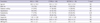
The baseline clinical characteristics are summarized in Table 1. Total 100 AMI (64 patients with STEMI) were enrolled in this study. Mean age of enrolled patients was 60.3 ± 13.3 yr. We compared all baseline clinical and laboratory data between STEMI and NSTEMI patients. There was no significant difference between two groups on the prevalence of diabetes mellitus (DM, 26.6% vs 38.9%, P = 0.201), hypertension (HTN, 50.0% vs 44.4%, P = 0.594), and smoking (62.5% vs 58.3%, P = 0.685). The prevalence of dyslipidemia was higher in the STEMI patients than in the NSTEMI patients (79.7% vs 55.6%, P = 0.01).
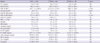
Mean serum Trx-1 level was 3.43 ± 1.80 ng/mL, initial CK level was 368.3 ± 531.3 IU/L, peak CK level was 1,951.7 ± 2,392.8 IU/L, peak CK-MB level was 217.8 ± 867.2 ng/mL, and cTnT level was 0.61 ± 1.6 ng/mL in all patients. There were no significant difference between STEMI and NSTEMI patients in serum Trx-1 level (3.44 ± 2.18 ng/mL vs 3.41 ± 0.80 ng/mL, P = 0.906), initial CK level (384.3 ± 607.5 IU/L vs 339.8 ± 364.4 IU/L, P = 0.690). But, STEMI group showed higher peak CK level (2,571.8 ± 2,754.5 IU/L vs 849.1 ± 764.6 IU/L, P < 0.001), and peak CK-MB level (207.5 ± 174.3 ng/mL vs 87.1 ± 96.2 ng/mL, P < 0.001). Mean number of WBC was 11.81 ± 4.64 × 10³/µL, uric acid was 5.12 ± 1.54 mg/dL, NT-proBNP was 2,021.8 ± 5,424.5 pg/mL, and fasting LDL-C was 112.35 ± 36.6 mg/dL in all patients (Table 2).
PrePCI TIMI flow grades of enrolled patients were grade 0 (n = 42), 1 (n = 6), 2 (n = 19), 3 (n = 33) and TIMI flow grade of postPCI were grade 0 (n = 1), 1 (n = 1), 2 (n = 11), 3 (n = 87). The composition of pre-and postPCI TIMI flow grade in the two types of AMI were comparable. Site of occlusion in AMI culprit LAD coronary artery was proximal (n = 45), middle (n = 53), and distal location (n = 2). Pain duration of all patients was 639 ± 2,078 min, STEMI patient showed a tendency of short pain duration compared to NSTEMI patients (304 ± 650 min vs 1,252 ± 3,326 min, P = 0.09).
Mean DTBT was 205 ± 360 min, and STEMI patients showed short DTBT compared to NSTEMI patient (57 ± 20 min vs 483 ± 508 min, P < 0.001). EF of left ventricle of AMI patients was 57.1 ± 15.7% (54.2 ± 16.8% in STEMI, 62.2 ± 11.9% in NSTEMI, P = 0.013), and WMSI of AMI patients was 1.37 ± 0.34 (1.46 ± 0.34 in STEMI, 1.22 ± 0.28 in NSTEMI, P = 0.001). Therefore, there were significant differences in DTBT, left ventricular EF and WMSI between two types of MI (Table 3).
The correlation between serum Trx-1 level and myocardial damage
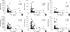
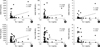
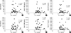
We analyzed correlation between baseline or peak cardiac enzyme and serum Trx-1 concentration. Trx-1 shows significant correlation with initial CK (P = 0.005, r = 0.281), initial cTnT (P < 0.001, r = 0.453), peak CK (P = 0.001, r = 0.316) in all patients (Fig. 1). In STEMI patients, there was also linear correlation between serum Trx-1 and initial CK (P = 0.008, r = 0.329), initial cTnT (P = 0.001, r = 0.498), and peak CK (P = 0.005, r = 0.349) (Fig. 2). No statistical correlation was observed between cardiac enzymes and serum Trx-1 level in NSTEMI patients (Fig. 3).
The correlation between serum Trx-1 level and biochemical markers
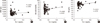
Trx-1 values did not have statistical correlation with BUN or hs-CRP. Serum uric acid (5.12 ± 1.54 mg/dL, P = 0.042, r = 0.210), and hemoglobin (14.0 ± 1.6 g/dL, P = 0.012, r = 0.251) have significant relation with Trx-1 level in all AMI patients. There is no correlation between WBC (11.81 ± 4.64×10³/µL, P = 0.674, r = 0.043) and Trx-1 level in all AMI patients (Fig. 4).
The correlation between serum Trx-1 level and clinical markers
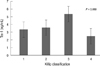
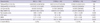
There was no statistical relationship between pre or postPCI TIMI flow grade and the serum Trx-1 level (prePCI: grade 0, 42%, 3.39 ± 1.18 ng/mL; grade 1, 6%, 3.12 ± 1.06 ng/mL; grade 2, 19%, 4.04 ± 2.73 ng/mL; grade 3, 33%, 3.19 ± 1.09 ng/mL, P = 0.406/post PCI: grade 0, 1%, 2.47 ng/mL; grade 1, 1%, 2.30 ng/mL; grade 2, 11%, 3.07 ± 0.99 ng/mL, grade 3, 87%, 3.50 ± 1.90 ng/mL, P = 0.745). There were no correlation between EF or WMSI and serum Trx-1 level (EF=57.1 ± 15.7%, P = 0.458, r = 0.075, WMSI=1.37 ± 0.34, P = 0.097, r = 0.167). And, serum Trx-1 value did not show statistical relationship with chest pain duration (P = 0.76), DTBT (P = 0.87 in all, P = 0.61 in STEMI, P = 0.80 in NSTEMI patients). However, there was tendency of relationship between serum Trx-1 level and the Killip classification (Killip I, 3.34 ± 1.61 ng/mL; Killip II, 3.58 ± 1.05 ng/mL; Killip III, 5.33 ± 4.57 ng/mL; Killip IV, 2.45 ± 0.29 ng/mL, P = 0.069) (Fig. 5). The Killip III population showed the highest value of serum Trx-1 level. Serum Trx-1 level did not have any relation for MACEs in enrolled AMI patients. MACEs occurred 2% in all AMI patients (1.6% in STEMI, 2.8% in NSTEMI), and there was no significant relation between Trx-1 value and incidence of MACEs (P = 0.78) (Table 3).
DISCUSSION
Our study provided evidences that the serum prePCI Trx-1 level showed good statistical relation with cardiac enzymes which are important markers for injured myocardial amount in STEMI, but not in NSTEMI. Trx was investigated in eukaryote level for several decades, but the clinical relevance of Trx was exploited in very recently (17). Clinically, the serum Trx level showed meaningful relationships with systolic dysfunction and symptom severity in heart failure (HF) and severity of ischemic heart disease (18). And, higher level of serum Trx means higher level of oxidative stress, overshooting of glucocorticoid system, and higher inflammatory status in HF patients (19, 20). Post-angioplastic serum Trx level showed inverse correlation with restenosis rate in peripheral arterial occlusive diseases in human (21). Pathological roles of Trx-1 in cardiovascular diseases were confirmed in various animal models. Trx-1 regulated angiotensin II-induced cardiac hypertrophy negatively through up-regulation of microRNA 98/let-7 family in murine model (22). And, the Trx downregulation exaggerated an angiotensin II infusion inducing cardiac hypertrophy in menopause rat (23). Trx-1 overexpressive transgenic mice showed better preservation of mitochondria and myofibril architecture, and better survival in adriamycin induced HF, and reduced the post-ischemic reperfusion injury of brain, especially in streptozotocin-induced diabetes mellitus animal model (7, 24). And, overexpression of Trx-1 limited CMCs apoptosis, preserved LV systolic function in animal models of MI (25, 26).
With these backgrounds, we investigated the correlation between serum Trx-1 levels with myocardial damage in AMI patients in this study. We analyzed 100 AMI patients retrospectively with culprit lesion in only LAD coronary artery, evaluated myocardial damage amount with various biochemical markers, and clinical variables including TIMI flow grade. We already have proven that prePCI Trx level had correlation with injured myocardial amount calculated by myocardial enzyme and WBC counts in AMI patients (10). However, this report enrolled relatively small number of patients (n=37), diverse characters and types of AMI patients in the aspects of pain duration, DTBT and culprit vessel location (10). AMI on left circumflex or right coronary artery can provoke extreme bradycardia and temporal hypotension related with right ventricular ischemia, so, these forms of AMI can show very wide range of oxidative stress and Trx-1 level. Therefore, we have focused on just LAD culprit AMI patients to make the homogeneity in anterior wall infarction with relatively large number and even character of patients (n=100), and the relationship among serum Trx level, myocardial damage amount, and TIMI flow grade in this analysis. We tried to confirm the pathological meaning of prePCI Trx level in relatively even condition of AMI patients, and we analyzed the difference of Trx level and its pathological meanings in STEMI and NSTEMI patients compared to our previous study (10).
There were linear correlations between serum Trx-1 level and several kinds of cardiac enzymes. In all AMI patients, the prePCI serum Trx-1 level showed good correlation with initial CK level (P = 0.005, r = 0.281), initial cTnT level (P < 0.001, r = 0.453), and peak CK level (P = 0.001, r = 0.316) (Fig. 1). In 64 STEMI patients, there was linear correlation between serum Trx-1 level and initial CK (P = 0.008, r = 0.329), initial cTnT level (P = 0.001, r = 0.498), peak CK level (P = 0.005, r = 0.349) (Fig. 2). These results strongly suggest that serum Trx-1 levels may reflect the extents of myocardial damage and oxidative stress in patients with STEMI. But, no correlation was observed between serum Trx-1 level and cardiac enzymes in patients with NSTEMI. So, we could speculate there were some different clinical meaning of Trx in STEMI and NSTEMI. According to the basic characteristics of two groups of AMI, STEMI showed narrow variation of pain duration and DTBT compared to NSTEMI. This uniformity and early hospital presentation of STEMI patient maybe contributes the meaningful relationship between prePCI Trx value and myocardial damage markers. And, relatively small number of patients and wide distribution of pain duration and DTBT in NSTEMI could be one cause for none of correlation between Trx-1 level and cardiac enzymes.
ROSs are very unstable molecules chemically, and half-life of them is very short. So, activities of antioxidative molecules also get fast change in cell or tissue level. Diverse stages of ischemia in NSTEMI also can be a cause for lack of relation with Trx-1 level to myocardial damage with this background.
Trx-1 level did not get statistical correlation with prePCI/postPCI TIMI flow grade, EF, WMSI, chest pain duration and DTBT. However, serum Trx-1 level had strong tendency of correlation with the Killip classification. The highest value of Trx-1 was observed in the Killip class III, not in class IV. We surmised that Trx-1 was severely exhausted in catastrophic situation with cardiogenic shock in the Killip IV, it might affect the lowest Trx-1 value in the Killip class IV. The level of prePCI Trx-1 in AMI patient might be closely related with myocardial damage and its related oxidative stress, not with pain duration or DTBT.
Extent of myocardial damage can be determined by complex summation composed of location of stenosis in culprit vessel, stenosis duration, stenosis severity (TIMI grade), presence of collateral flow, and myocardial susceptibility to ischemia. Therefore, just one or two clinical, biochemical, angiographic index cannot predict the extent of myocardial damage, and this complex theory will be the key of necessity to discover new myocardial damage extent marker, for example, Trx-1.
Although Trx-1 level had good correlation with several myocardial damage markers, the Trx-1 did not have the prognostic value for MACE in 6 months. It will be a profound limitation to use Trx-1 in prognosis marker in AMI. It involves consideration from various clinical aspects. We studied with relatively large number of patients to compare previous studies dealt with the relationship between clinical, biochemical findings with Trx-1. However, it will be still necessary much more number of patients to get the statistical meaning for the short, or mid-terms prognosis.
Unpredictably, prePCI serum Trx-1 level showed significant relationship with serum uric acid level and hemoglobin. Uric acid is byproduct of cellular metabolism and it reflects increased xanthine oxidase activity and oxidative stress. The xanthine oxidase system may also contribute to abnormal energy metabolism in human cardiomyopathy (27). And, uric acid level had association with microvascular disease, and it could determine coronary blood flow and patients prognosis especially in AMI (28). Also, hemoglobin has biological relationship with Trx-1, and oxidative stress. Hemoglobin undergoes oxidation-reduction reactions that lead to both generation and consumption of high amount of ROSs (29, 30). Furthermore, considerable amount of Trx-1 exist in RBC (29). So, even the meticulous separation of serum from whole blood, there is the possibility of RBC contamination into serum. With these backgrounds, the statistical correlation between Trx-1 and hemoglobin level can be explained.
This study has several limitations. We evaluated correlation between serum Trx-1 level and the data of patients of only AMI. We did not get any data about the relation between Trx-1 and angina- or ischemia-free patients. And, we checked just one time point of Trx-1 in prePCI, and we did not check the Trx-1 level evolution during admission. So, we could not show the Trx-1 evolution and its clinical meaning for postPCI period. Relatively small number of NSTEMI can be important hurdles to generalize the clinical meaning of Trx-1 into all AMI. This analysis needs a large scale, prospective study to form conclusive results, and give predictive power as a single biochemical maker of Trx-1 for AMI. And, fine methods for myocardial damage amount measurement, for example, myocardial magnetic resonance image, should be introduced to check the reliability of Trx-1 in the future.
In conclusion, for AMI patients with culprit lesion on only LAD coronary artery who were treated with PCI as a primary treatment, we investigated clinical relationship between prePCI serum Trx-1 level and myocardial damage. Results showed that prePCI serum Trx-1 level is associated with myocardial damage, which was measured by the myocardial enzyme. We can consider serum Trx-1 levels on prePCI as a predictor of myocardial injury in STEMI patients, not in NSTEMI patients.
 XML Download