

 PDF
PDF ePub
ePub Citation
 Print
Print


INTRODUCTION
Takotsubo cardiomyopathy (TC) is an increasingly reported syndrome characterized by transient apical left ventricular dysfunction in absence of low limiting coronary artery disease. Prolongation of QTc interval associated with TC has been previously reported in published case series. Bradycardia induced QT interval prolongation seemed to be amplified by the occurrence of TC, resulting in torsade de pointes. Temporary ventricular pacing at a high rate decreased the QT interval and prevented the recurrence of torsade de pointes. We report a case of torsade de pointes associated with QT prolongation and TC. Because QT prolongation and bradycardia persisted even after the resolution of TC, the patient received a permanent pacemaker.
CASE DESCRIPTION
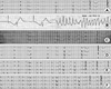
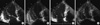
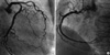
A 78-yr-old female with a medical history of hypertension was admitted to our hospital due to syncope on 20 July, 2010. Her antihypertensive medications were nifedipine (30 mg) and valsartan (80 mg). Upon arrival to hospital, the patient was fully conscious and alert with a pulse rate of 40 beats per minute and a blood pressure of 140/80 mm Hg. She had no chest pain but had orthopnea. No neurological deficit was noted. Brain CT and chest radiography showed normal findings. She had suffered from emotional stress because her grandson ran away from home. Electrocardiography (ECG) on admission showed an escape junctional rhythm of 35 beats per minute combined with T wave inversion in precordial leads and QT prolongation measuring 580 ms in the QTc interval (Fig. 1A). The serum levels of potassium, magnesium, and ionized calcium were within normal limits. The serum troponin T level was slightly elevated to 0.081 ng/mL (0-0.1 ng/mL). Echocardiography showed akinesia of mid and apical left ventricular (LV) walls with the systolic ballooning of the ventricular apex; LV ejection fraction was estimated to be 35% by a modified Simpson's method (Fig. 2A, B). Coronary angiography was performed for further evaluation of ischemic heart disease. This angiography demonstrated no significant coronary artery disease (Fig. 3). The ergonovine coronary spasm test showed negative findings. She was diagnosed with takotsubo cardiomyopathy based on echocardiographic and coronary angiographic findings. On the second day, the patient had sudden loss of consciousness and shock. ECG showed severe bradycardia (heart rate 20 beats per minute) with marked prolongation of QT (QTc = 720 ms) followed by torsade de pointes (TdP) (Fig. 1B). The patient was successfully resuscitated with DC cardioversion and intravenous magnesium sulfate. After temporary ventricular pacing, which was performed at a rate of 80 beats/min, the QTc interval decreased to 480 ms. On the fourth day, the temporary pacemaker was removed. At that time, ECG showed a marked bradycardia (heart rate of 40 beats per minute) with QT prolongation and R on T phenomenon (Fig. 1C). On the 10th day, we checked follow-up echocardiography, which showed normal LV systolic function without previous wall motion abnormalities (Fig. 2C, D). Also, the troponin T level normalized. ECG still showed a severe bradycardia with QT prolongation while we checked daily serial ECG up to the 20th day (Fig. 1D). Therefore, we planned to insert a permanent pacemaker to prevent recurrent TdP, although LV systolic dysfunction of the patient was recovered. On the 21st day, she received a dual-chamber pacemaker. Since then, additional event did not occur and the patient was discharged. One and 3 months after the index procedure of permanent pacemaker insertion, ECG still showed QT prolongation (QTc = 552 ms) with T-wave inversion at precordial leads (Fig. 1E, F).
DISCUSSION
TdP denotes a polymorphic ventricular tachycardia associated with long QT syndrome, and is a life-threatening arrhythmia. QT prolongation is the surface ECG manifestation of abnormal repolarization of myocardial cells due to problems with cellular ion channels. The disorder is classified as either congenital or acquired. Acquired QT prolongation may be associated with the following conditions: 1.electrolyte depletion (especially potassium or magnesium) 2. drugs that affect myocardial ion channels 3. structural heart disease. Also, the association of TC with Tdp has rarely been reported (1, 2).
The relationship between TC and abnormal repolarization has been well documented. Patients with TC generally show ST elevation, T-wave inversion, Q-wave formation and QTc interval prolongation in ECG. The prevalence of QTc interval prolongation among TC patients is high, ranging from 50% to 100% according to different case series. This is probably because systolic dysfunction is associated with both TC and QT-interval prolongation (3). Although QT-interval prolongation is prevalent among TC patients and might precede TdP, the later has rarely been reported in TC patients. Danney et al. (5) first reported TdP occurring in patients with TC. Since then, numerous reports of TdP related to TC have been published (4, 5). Also, bradycardia with or without AV block and suspicious of congenital long QT syndrome are the risk factors of TC-associated TdT (4). However, permanent pacemaker insertion has been performed on a few patients (6, 9). In general, QT interval prolongation and ST-T changes occur during the acute or subacute phase of illness, and these ECG changes typically normalize within several weeks of improvement in LV wall motion (3).
In our case, QT prolongation and ST-T change persisted for 3 months. Inoue et al. (6) reported a patient who underwent permanent pacemaker insertion due to complete AV block associated with TC. In their case, the ECG of the patient showed complete AV block even after recovery from illness. Mahida et al. (7) and Sasaki et al. (8) also reported 2 cases of suspicious congenital long QT syndrome associated with TC. During the previous regular medical check up of the present case, the ECG of the patient have shown normal findings. After development of her illness, QTc interval didn't improved, even after normalization of LV wall motion and cardiac enzyme level. Based on her previous medical check up ECG and history, a diagnosis of acquired long QT syndrome associated with TC was felt to be likely. And, we inserted a permanent pacemaker because sinus bradycardia (40 beats per minute) and QT prolongation (QTc = 550 ms) persisted for 2 weeks even after recovery from LV dysfunction. To date, the association between TC, bradycardia and TdP is not exactly known. After recovery from LV dysfunction in TC, QT prolongation usually normalizes. Kurisu et al. (9) stated that prolongation of bradycardia-induced QT interval may be augmented by the occurrence of takotsubo cardiomyopathy, resulting in TdP. In their cases, permanent pacemaker insertion was also performed because bradycardia and complete AV block persisted after the recovery from LV dysfunction. Although TdP has rarely been reported in patients with TC, the clear relationship between the 2 conditions has yet to be determined. However, QT prolongation and bradycardia could be persistent even after full recovery of TC, and permanent pacemaker insertion may be a treatment option of long QT syndrome related with TC.
 XML Download