

 PDF
PDF ePub
ePub Citation
 Print
Print


INTRODUCTION
The proportion of candidemia cases caused by non-albicans Candida species has been increasing (1). Some non-albicans Candida species are associated with resistance to the usual antifungal azoles (2). C. haemulonii, a non-albicans Candida species, does not frequently cause human infections (3). Microbiologically, the susceptibility profile of C. haemulonii shows that it is resistant to amphotericin B and other antifungal agents such as azoles (4), which have often been associated with clinical treatment failure (5, 6). No treatment regimen for invasive C. haemulonii infections has been clearly established.
We investigated a catheter-related candidemia infection due to C. haemulonii and report on resolution of the infection using caspofungin, an echinocandin antifungal agent.
CASE DESCRIPTION
On June 25, 2009, a 67-yr-old man with a cerebral infarction was hospitalized in the department of rehabilitation medicine for physical rehabilitation. Prior to admission, he experienced cerebral infarction and was hospitalized for two months, beginning in March, 2009. He had received a tracheostomy at his previous admission.
A physical examination did not reveal any signs of infection, including at the tracheostomy site. The patient received a right subclavian venous catheter for total parenteral nutrition on the second day of hospitalization because of the risk of aspiration when receiving nasogastric tube feedings.
On August 15 (the 50th day of hospitalization), the patient had a temperature of 37.8℃, a white cell count of 10.4 × 10⁹/liter, and a C-reactive protein level of 10.8 mg/liter. However, the blood culture failed to reveal any infectious organism. The mild fever continued, and the level of CRP and white cell count continued to increase.
On August 29 (the 64th day of hospitalization), a blood culture collected 72 hr previously showed yeast growth, and the subclavian venous catheter was removed. Intravenous fluconazole (400 mg once a day) was administered as empirical therapy, pending identification of the yeast. A qualitative culture of the catheter tip also produced a yeast culture.
The yeast from the blood culture and catheter tip, designated as LYS-1, was identified as Candida sp. on the seventh day of fluconazole therapy. The physician continued the fluconazole therapy for the candidemia. The blood cultures performed on the fourth and 14th days of fluconazole therapy were still positive for yeast, and the mild fever persisted.
On September 15 (the 18th day of fluconazole treatment), the infectious disease department was consulted and recommended an echocardiography, a bone scan, an abdominal CT, and Doppler sonography for venous thrombosis. The echocardiograph revealed no signs of infectious endocarditis. The bone scan, abdominal CT, and Doppler sonography revealed no significant findings.
On September 17 (the 84th day of hospitalization), the treatment regimen was changed to caspofungin (50 mg daily after a 70 mg loading dose). The blood culture was sterile on the third and tenth days of caspofungin therapy. On the second day of the caspofungin treatment, the patient became afebrile and showed clinical improvement. In resolution, the patient underwent a 17-day course of caspofungin and remained stable after discontinuation of the antifungal agent.
Molecular identification
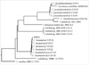
Conventional automated methods in the clinical microbiology laboratory identified the LSY-1 isolate as Candida sp. Thus, we attempted to identify the culture at the species level using a molecular method. To identify the isolate LSY-1, a portion of the large subunit (LSU) rRNA gene was amplified using the primers LSU-F (5'-GCATATCAATAAGCGGAGGAAAAG-3') and LSU-R (5'-GGTCCGTGTTTCAAGACG-3'). Template DNA and 20 pM of each primer were added to a PCR mixture tube (Accu-Power PCR PreMix; Bioneer, Daejeon, Korea). The reaction mixture was then subjected to 35 cycles of amplification. Each cycle consisted of 30 sec at 95℃, 30 sec at 50℃, and 1 min at 72℃, followed by a final extension at 72℃ for 1 min. The amplified PCR product was purified for sequencing using a PCR purification kit. The purified PCR product was sequenced directly using the same primers as those used in the PCR amplification. The determined sequences (519 bp) were compared with the Gen-Bank public database using the BLASTn program (http://blast.ncbi.nlm.nih.gov/Blast.cgi). The LSU rRNA gene sequence of the isolate LSY-1 was a complete match with the corresponding sequence of the reference strain, C. haemulonii strain TJY2d (GenBank accession numbers EU359820). In addition, the isolate in question showed many similarities to the sequences of the C. haemulonii strains and similarities of less than 85% with other Candida species (Fig. 1). Thus, the sample was confidently identified as C. haemulonii.
DISCUSSION
C. haemulonii was originally described from a sample taken from the gut of a blue-striped grunt (Haemulon scirus) in 1962 (7), and Lavarde et al. (8) reported the first clinical isolation of this fungus from a human in 1984.
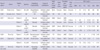
Several meaningful cases of C. haemulonii candidemia have been reported (Table 1). Antifungal resistance is the focus of attention in the management of invasive candidiasis. In previous cases, most C. haemulonii were resistant to both fluconazole and amphotericin B (5, 9). Clinically, when amphotericin B was administered empirically, it failed to eradicate C. haemulonii candidemia (5). The majority of cases in which fluconazole was administered empirically also failed to eradicate candidemia (5, 6). Although fluconazole administration has been shown to resolve candidemia, these cases could be considered transient candidemia combined with the removal of the intravenous catheter. Therefore, the resistance of C. haemulonii poses a therapeutic challenge for the treatment of invasive candidiasis.
Microbiologically, previous reports and our case show that C. haemulonii is susceptible to echinocandins such as caspofungin or micafungin (5, 6, 10), and that it was not resistant to new triazoles such as voriconazole. However, it is not certain that echinocandins or voriconazole are actually efficacious in the treatment of C. haemulonii candidemia. There have been only two cases of clinical success in the eradication of C. haemulonii candidemia via echinocandins administration (5, 6). One case used micafungin, and the other used caspofungin. Of those, in the treatment with caspofungin of a neonate, echinocandin was combined with amphotericin B. Therefore, our case could demonstrate clinical success with caspofungin administration in the eradication of C. haemulonii candidemia in an adult patient.
In our case, the VITEK II (bioMérieux SA) clinical yeast identification system was used for initial identification of C. haemulonii, and sequence analysis of the partial 26s rRNA gene (519 bp) was performed for confirmation. The 519-bp sequences of the isolate matched completely with the corresponding sequences of the reference strain, C. haemulonii strain TJY2d, available in the GenBank database (accession number EU359820). In recent studies, the identification results of the VITEK 2 system have closely corresponded with those of molecular methods for the identification of C. haemulonii, while the VITEK 1 system and the API 32C system usually fail to identify C. haemulonii (5, 10).
Known risk factors for candidemia are use of central-venous catheterization, total parenteral nutrition, previous multiple antibiotics, previous steroid therapy, previous abdominal surgery, and an immunocompromised status (11, 12).
In our case, long-term central-venous catheterization for hyperalimentation was the associated risk factor for candidemia, and no other source of infection suggested the possibility of catheter-related candidemia. In previous case reports, the majority of patients also received an intravenous central line and were in an immunocompromised status.
Considering the recent case series of invasive C. haemulonii infections, C. haemulonii is considered to be an emerging yeast pathogen for which the optimal strategy of patient management has yet to be elucidated.
In conclusion, as found in the previous case reports, fluconazole and amphotericin B are not reliable empirical antifungal agents for the treatment of C. haemulonii candidemia. Echinocandins, such as caspofungin or micafungin, may be an appropriate empirical choice of antifungal agent for invasive C. haemulonii infections. New triazoles require additional clinical testing for treatment of C. haemulonii infection.
 XML Download