

 PDF
PDF ePub
ePub Citation
 Print
Print


INTRODUCTION
During the spring of 2009, the Centers for Disease Control and Prevention (CDC) confirmed the first two cases of human infection with the 2009 influenza A (H1N1) virus in the USA (1). As the virus spread rapidly to other regions of the world, the World Health Organization (WHO) declared the first phase 6 global influenza pandemic of the century on 11 June 2009 (2). By 23 December 2009, there were > 11,516 deaths worldwide; however, the reported number of fatal cases is an under-representation of the actual number as many deaths are never tested or recognized as influenza-related (3).
In Korea, the first confirmed case of H1N1 influenza was reported on 1 May 2009 and the first fatal case was documented on 15 August 2009. Until 31 January 2010, 740,835 patients were confirmed with pandemic H1N1 2009 and 225 of them were reported to have died. (4). In spite of various reports of critical illness caused by the 2009 H1N1 influenza in North America, Europe, and Oceania, there have been no reports focusing on deaths in Asia (5-10). The current report describes the clinical and epidemiologic characteristics of the first 115 deaths associated with the 2009 pandemic influenza A (H1N1) virus infection in Korea between 15 August and 26 November 2009.
MATERIALS AND METHODS
Prior to the H1N1 epidemic, Korea had a passive influenza surveillance system. Every week, 820 private medical institutions reported patients with influenza-like illnesses (ILIs) and 105 private medical institutions updated respiratory viral infections by laboratory tests.
From late June 2009, the Korea Centers for Disease Control and Prevention (KCDC) began active mortality surveillance in inpatients within nationwide community-based hospitals, as determined by the government. A fatal case was defined as a person with a confirmed H1N1 influenza 2009 infection based on ante- or post-mortem specimens who died from a clinically compatible illness or complications attributable to that infection. There should be no period of complete recovery between illness and death, and no alternative agreed upon cause of death.
With the first fatal case report of the 2009 pandemic influenza A (H1N1) virus on 15 August, we collected several types of data for the fatal cases using a standardized format that included demographic features, underlying diseases, initial symptoms, warning signs, chest radiographs, laboratory findings, complications, and treatment course. Data were obtained by physicians (Epidemic Intelligence Service Officers) based on a review of medical records and discussions with the patient's attending physician. All laboratory findings were clinically driven and recorded during the hospitalization, regardless of the type of ward the patient was admitted to.
The body mass index (BMI; kg/m2) was calculated for patients in whom the height and weight were available to determine whether or not the patient was obese. Obesity was defined as a BMI ≥ 25 kg/m2 in adults ≥ 18 yr of age, as based on WHO Asia-Pacific diagnostic criteria (11).
All analyses were carried out with the use of SPSS 12.0K.
RESULTS
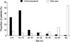
The median age of the dead patients was 61 yr (interquartile range [IQR], 0.2-97 yr). Fifty-eight patients (50.4%) were females, 16 patients were < 20 yr of age, and most of the patients (53.9%) were > 60 yr of age. In contrast, the majority of confirmed patients (78.1%) were < 20 yr of age and only 1.2% of the patients were > 60 yr of age (Fig. 1).
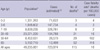
A total of 723,974 people in Korea had ILIs based on positive reverse transcriptase polymerase chain reaction (RT-PCR) during the study period. With this denominator, the ILI mortality rate was 16 deaths per 100,000 cases. The age-specific mortality rate had a J-shaped curve (Table 1).
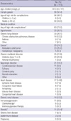
Fifty-six patients (48.7%) were at high risk based on age (< 5 yr or ≥ 65 yr), and 83 patients (72.2%) had 1 or more underlying medical conditions. Ninety-six patients (83.5%) had a high-risk condition (age or underlying disease). A high proportion of children (64.3% of 0-17 yr of age) had no documented underlying disease, while 86% of people > 65 yr of age had an underlying disease. The most frequently identified underlying medical condition was chronic lung disease in 30 patients (26.1%). Other underlying conditions included cancers, diabetes, neurologic disorders, cardiac disease, chronic renal disease, chronic liver disease, and immunosuppressed conditions. Height and weight were available for 47 of 115 patients (40.9%) who were > 18 yr of age. Of the 47 patients, 11 (23.4%) were obese (BMI ≥ 25 kg/m2). There were no deaths in pregnant woman (Table 2).
Diagnostic findings
Evidence of concurrent bacterial infections was found in sputum or blood specimens from 34 patients (29.6%). Of these 34 patients, 28 had confirmed bacterial infections in sputum specimens. The most common bacteria isolated in the sputum specimens were Staphylococcus aureus and Klebsiella pneumoniae. These infections were documented in eight patients each. One-half of S. aureus isolates were methicillin-resistant (MRSA). Based on bronchoalveolar lavage (BAL) specimens, five patients had Acinetobacter baumannii, four patients had Candida albicans, three patients had Streptococcus pneumonia, two patients had Pseudomonas aeruginosa, and three patients had Aspergillus fumigatus. In addition, sputum positive for acid-fast bacilli, Escherichia coli, and Pneumocystis jiroveci based on BAL specimens were demonstrated in one patient each. Although different from the sputum culture, a 7-yr-old child was seropositive for Mycoplasma IgM and had a 4-fold rise in serial titers for anti-Mycoplasma IgG.
Bacterial organisms were cultured in blood specimens from 10 patients. Three patients had Klebsiella pneumoniae, one patient had C. albicans, and one patient each had vancomycin-resistant Enterococcus, Enterobacter, Pseudomonas aeruginosa, Streptococcus pneumoniae, S. capitis, and yeast. In addition, Citrobacter braakii and E. coli were isolated from ascites and urine.
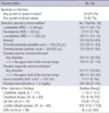
Leukocytosis and thrombocytopenia occurred frequently in patients with ILIs. Anemia and abnormal liver function tests was also common. Six patients who had no evidence of hepatitis B or C virus infections were thought to have severe acute hepatitis. The erythrocyte sedimentation rate (ESR), C-reactive protein (CRP), creatine kinase (CK), and lactate dehydrogenase (LDH) levels were increased in patients with ILIs (Table 3).
Complications
Of the 113 patients who underwent chest radiography on admission, 97 patients (85.8%) had findings that were consistent with pneumonia. Pneumothoraces occurred in two patients after central line insertion and ventilator care. The most common cardiac complication was myocarditis (13 patients [11.3%]). Pulmonary emboli were detected in two patients by CT or echocardiography and blood testing. Pericarditis was documented in two patients. Myocardial infarctions were noted in 2 patients; 1 patient had angina with 3-vessel disease as an underlying condition and the other patient was a smoker without other underlying diseases. In addition, a ruptured abdominal aortic aneurysm was founded.
Seven patients (6.1%) had encephalopathy based on EEG, brain MRI, or brain CT; 2 of the patients were shown to have the 2009 pandemic influenza (H1N1) virus by RT-PCR of cerebrospinial fluid. Except these patients, intracranial hemorrhage and acute cerebral infarction were noted in 1 patient each.
Five patients (4.3%) had gastrointestinal bleeding; 2 of the patients had colon cancer and the others had no definite underlying disease to account for the bleeding. Intestinal obstruction occurred in three patients; one of the patients had a history of intestinal surgery. Among other two patients with peritonitis, one patient had pancreatic cancer and one patient had no underlying disease. A liver abscess was documented in one patient with diabetes and stroke.
Eighteen patients (15.7%) who had no underlying chronic renal disease were suspected to have acute renal failure; 9 of the patients were treated with dialysis, continuous renal replacement therapy, or diuretics, and the other patient had elevated creatinine levels (> 2.0 mg/dL). Rhabdomyolysis was noted in 4 patients (3.5%) who had a CK level > 3,000 U/L (10 times the normal upper range in adults); 1 patient had a CK level > 30,000 U/L.
Anti-viral treatment
Of the 115 patients, 100 (87%) received anti-viral drugs. Of these 100 patients, 98 received oseltamivir, 1 received zanamivir, and 1 received combination therapy with oseltamivir plus zanamivir. All of the patients who were treated with oseltamivir received the recommended dose that is approved by the US Food and Drug Administration, but 15 adult patients were treated with high-dose oseltamivir (300 mg a day). The median time from the onset of illness to the initiation of anti-viral therapy was 3 days (IQR, 0-21 days); 36% of these patients received anti-viral therapy within 48 hr after the onset of symptoms.
Clinical course
The median time from symptom onset to death was 8 days (IQR, 1-33 days). Fifteen patients died within 48 hr since the onset of symptoms and 4 patients died within 24 hr.
We evaluated 63 patients who were admitted to the intensive care unit (ICU), except confirmed cases during hospitalization for other diseases and cases with unknown symptom onset. The median time from symptom onset to hospital admission was 2 days (IQR, 0-22 days), the median time from hospitalization to ICU admission was 0 days (IQR, 0-17 days), and the median time from ICU admission to death was 4 days (IQR, 0-26 days).
DISCUSSION
Our data suggest that age ≥ 65 yr has the lowest incidence and the highest case fatality rate for the 2009 influenza A (H1N1) virus. Our age-specific case fatality rates follow a pattern similar to the J-shaped distribution described in the Mexican, English, and Japanese studies of the current pandemic (13-15). The incline of the curve became steeper with older age older. The median age of our analysis was much older than the median age reported in other countries (median age range, 37-53 yr) (7-9, 16). These results differ from the early 2009 pandemic in which there was a high case fatality occurring among young healthy adults (7, 17). The case fatality of our study (16 per 100,000) is lower than that of previous report (30 per 100,000) based on the confirmed patients in Korea (4). This is because not all patients with ILI were confirmed by RT-PCR.
The finding that the majority of deaths occurred in older patients explains that the proportion of patients who had gastrointestinal symptoms, headaches, and myalgias (systemic symptoms) at the onset of illness was much lower than previous reports (5, 8, 9). Generally, the younger age group is more likely to have systemic symptoms in seasonal influenza infections (18). There were no pregnant women enrolled in our study.
There are a greater number of cancer patients (24.3%) with underlying medical conditions compared to previous studies (range, 2.6%-7.9%) (7, 14). Interestingly, cancer patients are infected more often during hospitalization to manage other medical diseases (P = 0.005).
Among critically ill patients with severe 2009 influenza A (H1N1) infections, obesity was more common than in the general population; however, increased BMI has not emerged as a predictor of mortality (8, 9). Although data regarding height and weight was available for only 47 adult patients (40.1%) in our study, 11 of these patients (23.4%) were obese based on the Asia-Pacific definition of obesity (11). The prevalence of obesity in our study was much lower than in the general adult Korean population (31.7%) (19).
Even though all of the patients were not evaluated with sputum cultures, evidence of concurrent bacterial co-infections in sputum specimens was shown in 29.6% of 115 fatal cases. The result was similar to the report in the USA in which a 29% bacterial co-infection rate was shown in lung tissue specimens from 77 deaths (20). The most common pathogens recovered were Klebsiella pneumoniae (8 patients) and S. aureus (8 patients) in our study, compared to Streptococcus pneumoniae (10/77) in the USA.
The data of our study were derived from the KCDC, which had a detailed description of deaths associated with the 2009 influenza A (H1N1), and differed with respect to race based on a previously published report. These observations of the epidemiologic risk factors and typical clinical features of fatal cases will help in the diagnosis and clinical management of severely ill patients with pandemic influenza A (H1N1).
This study had several limitations. First, we evaluated only fatal cases with confirmed 2009 H1N1 infections, so a comparative study of survivors and non-survivors could not be conducted. The group in our study may not be representative of those who were not tested. Second, our data were gathered while the epidemic was ongoing in Korea; the findings after the end of the pandemic may be different owing to the effect of massive vaccination. Third, the absolute low number of deaths in the very young age group could lead to a different interpretation. Finally, it is impossible to compare our case fatality rate to that of other countries directly because every country has a different calculation method of the estimated cases.
In Korea, from the end of October, anti-viral medications were administered to patients who met the definition of an ILI with or without laboratory confirmation. As of 31 December, approximately 9 million people have been vaccinated, including students in elementary, middle, and high schools since early November, through the national pandemic influenza H1N1 vaccination program. As a result, the trend of ILIs and deaths decreased rapidly. Considering the second wave of the outbreak in the spring of 2010, we have to make a vaccination plan for the older age group with underlying disease and prevent known complications. If possible, we expect to understand the 2009 H1N1 virus and manage patients appropriately by performing a mathematically well-designed mortality study and a comparative study involving survivors versus non-survivors.
 XML Download