

 PDF
PDF ePub
ePub Citation
 Print
Print


INTRODUCTION
Clonorchiasis is a trematodiasis caused by the Chinese liver fluke which is transmitted by snails. It is generally acquired by eating raw, inadequately cooked, or the picked flesh of freshwater fish. The geographic distribution of clonorchiasis is largely confined to the South East Asia including Korea, China, Hong Kong, Japan, Taiwan, and Vietnam (1-3). With the increasing popularity of travel to these countries as well as the global migration of Asians, physicians need to be aware of the condition. In Korea, through the high level growth of the economy, infection rates of soil-transmitted helminthiases have reduced remarkably. However, a recent survey revealed that snail-transmitted trematodiasis, in particular Clonorchis sinensis, remains highly prevalent, especially in the riverside areas of Korea (2, 3). The adult flukes take up residence most commonly in the medium and small intrahepatic bile ducts, and occasionally in the gallbladder, pancreatic duct and extrahepatic bile ducts. The flukes along with desquamated epithelial cells and metabolites, cause mechanical obstruction of the biliary tract, leading to inflammation and periductal fibrosis, which results in recurrent pyogenic cholangitis (4, 5). In this report, we present a rare case of Clonorchis sinensis infection with obstructive jaundice caused by duodenal papillitis alone without mechanical bile duct obstruction, which was treated by endoscopic sphincterotomy and oral praziquantel.
CASE DESCRIPTION
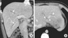
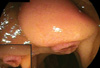
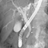
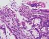
A previously healthy 26-yr-old male was referred to our hospital because of recently recognized jaundice on May 22, 2008. He complained of anorexia, fatigue, and weight loss of 8 kg over the previous eight months. He lives in the suburbs of Seoul. The medical history was unremarkable except for ingestion of raw freshwater fish. The patient had a blood pressure of 124/81 mmHg, a heart rate of 80 beats/min, a respiration rate of 15 breaths/min, and a body temperature of 36.7℃. The physical examination was unremarkable except for generalized jaundice. The laboratory data revealed white blood cell count 7,100/µL (normal range 4,000-10,000/µL) with 18.3% eosinophils (0-4%), hemoglobin level 12.4 g/dL (13-17 g/dL), platelet count 231,000/µL (130,000-350,000/µL), total bilirubin 11.3 mg/dL (0.22-1.2 mg/dL), direct bilirubin 8.7 mg/dL, aspartate aminotransferase 58 IU/L (< 40 IU/L), alanine aminotransferase 248 IU/L (< 40 IU/L), alkaline phosphatase 563 IU/L (66-220 IU/L), gamma glutamyltransferase 265 IU/L (8-66 IU/L), and amylase 86 U/L (28-100 U/L). Serologic testing of viral markers showed HBs Ag (-), antiHBs Ab (+), antiHAV IgM (-), and HCV Ab (-). The abdominal CT scan showed mild dilatation of the intrahepatic bile ducts (Fig. 1). Because Korea is an endemic area for C. sinensis infection and the patient had a history of ingestion of raw freshwater fish as well as peripheral eosinophilia, we initially suspected biliary obstruction due to C. sinensis infection. Therefore, endoscopic retrograde cholangiopancreatography (ERCP) was planned. On duodenoscopy, the duodenal papilla showed an edematous and bulging configuration with marked hyperemic changes at the orifice (Fig. 2). Cholangiography showed a mild degree of bile duct dilatation with ductal wall irregularities and indentations. However, there was no definite bile duct stricture or intraluminal filling defects, including stones or parasitic adult worms, noted (Fig. 3). We performed endoscopic sphincterotomy for effective bile drainage through the duodenal papilla and sweeping of the bile duct by a balloon catheter. However, no stones or adult worms were identified. We next performed multiple forceps biopsies at the duodenal papilla and inserted a nasobiliary drainage tube up to the second branch of the intrahepatic bile duct for bile collection. Pathology examination of the duodenal papilla showed chronic active inflammation with many eosinophilic cells in the mucosal layer (Fig. 4). In addition, there were parasitic eggs, consistent with C. sinensis found in the drained bile fluid (Fig. 5). Follow up liver function testing five days after the ERCP was dramatically improved: total bilirubin 4.7 mg/dL, aspartate aminotransferase 108 IU/L, alanine aminotransferase 224 IU/L, alkaline phosphatase 336 IU/L, and gamma glutamyltransferase 122 IU/L. The patient was discharged with oral praziquantel (25 mg/kg three times a day) seven days after the ERCP. Two months later after discharge from the hospital, the patient returned for follow up; he was in healthy condition, recovered his body weight and all laboratory studies had returned to the normal range.
DISCUSSION
Hepatobiliary complications of clonorchiasis include cholelithiasis, pyogenic cholangitis, cholecystitis, biliary obstruction, and cholangiocarcinoma secondary to mechanical injury to the biliary epithelium by the suckers of the worm and prolonged inflammation (4-6). Thus, most patients with complications of chronic C. sinensis infection present with acute cholangitis due to biliary obstruction by the adult worms or stones. However, painless jaundice is usually due to a bile duct stricture, benign or malignant. In our case, the patient presented with painless jaundice and a long history of weight loss. This presentation was suggestive of a malignant biliary obstruction. However, the frequent ingestion of raw fish over a long time, and the eosinophilia in the peripheral blood, suggested that C. sinensis related extrahepatic biliary stricture be included in the differential diagnosis. However, bile duct stricture was not present on ERCP. Instead, bile duct dilatation with irregularities and indentations of the ductal wall were present, which suggested a long history of C. sinensis infection (7, 8). Furthermore, a definite duodenal papillitis, sufficient to cause an obstructive jaundice, was found and confirmed histopathologically.
Duodenal papillitis is an inflammatory disorder involving the mucosa overlying the major duodenal papilla. It may be a reflection of an underlying biliary or pancreatic disorder associated with a clinically acute inflammatory condition such as acute cholangitis, acute pancreatitis or acute exacerbation of a chronic pancreatitis (9). Most inflammatory cells, in a biopsy specimen of the papillitis, are monocytes, and frequently associated with various degrees of neutrophils according to the severity of papillitis. In our case, the main inflammatory cell infiltrate in the mucosa were eosinophils. The possible mechanism of the heavy eosinophilic infiltration might be an allergic reaction to the metabolites released from the adult C. sinensis worms in the bile duct (10). This finding along with evidence of C. sinensis eggs in the drained bile fluid and the cholangiographic findings supported a parasitic infection.
The therapeutic role of endoscopy has not been fully evaluated in C. sinensis infection because most patients are treated by oral praziquantel treatment alone even if many adult worms were present in the bile duct (11). We could not find any other causes of the obstructive jaundice, including stones, parasitic worms, biliary stricture, or mass. C. sinensis related duodenal papillitis was the only possible explanation of the obstructive jaundice. Therefore we performed endoscopic sphincterotomy prior to oral praziquantel treatment for facilitating relief of biliary ductal pressure due to duodenal papillitis. Fortunately the patient's jaundice after the endoscopic sphincterotomy was dramatically relieved.
In conclusion, duodenal papillitis causing obstructive jaundice is a rare presentation of C. sinensis infection. Endoscopic sphincterotomy might be an effective therapeutic tool to relieve obstructive jaundice in duodenal papillitis due to C. sinensis infection.
 XML Download