

 PDF
PDF ePub
ePub Citation
 Print
Print


INTRODUCTION
Dopa-responsive dystonia (DRD) is a progressive primary dystonia that is characterized by onset during childhood, circadian fluctuation of symptoms and a dramatic and sustained response to low doses of oral administration of levodopa (1). DRD is frequently caused by GTP-cyclohydrolase 1 (GTPCH1) deficiency that up to 87% of the DRD is caused by mutations in the GCH1 gene encoding GTPCH1 (2, 3).
Although it is well-known that the clinical features of GTPCH1-deficiency can be extremely variable including benign adult-onset parkinsonism, various types of focal dystonia, DRD simulating cerebral palsy or spastic paraplegia, clinical diagnosis is still a challenge in some instances (4).
CASE DESCRIPTION
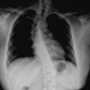
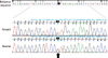
She was referred to our clinic when she was 30 on February 22, 2009. It resulted in frequent falls. Her symptoms were mild in the morning and more prominent toward the end of the day. As the disease progressed, she had pes equinovarus posture at rest in both feet. At the age 15 she needed the support of two people to walk a short distance and started to use a wheelchair. At this age she also experienced stiffness and twisting in upper limbs, neck, and trunk. She found it difficult to write and use a chopstick. Her birth and developmental history was unremarkable. She had no prior history of head injury, meningitis, encephalitis, or febrile seizures. There was no known history of motor disorder in her family. She was initially diagnosed as having cerebral palsy. Brain CT was normal. She received physiotherapy and muscle relaxants with no benefit. When she was 20, Madopar® 250 mg was prescribed by neurologist. Her symptoms dramatically improved within days, and she could walk independently. She was referred to our clinic when she was 30. She had been taking levodopa 200 mg/day for 10 yr with sustained benefit without emergence of motor fluctuations or other neurologic manifestations. She felt well and was leading a normal life. The fixed scoliotic deformity of thoracolumbar spine was noted on chest radiogrdaphy (Fig. 1). Genetic testing using direct sequencing revealed a novel initiation codon mutation (c.1A>T; p.Met1Leu) in the GCH1 gene (Fig. 2).
DISCUSSION
DRD-causing mutations in GCH1 include point mutations, small insertions, deletions and whole exon deletions (2, 11). We identified a novel mutation in the initiation codon. We speculate that the GCH1 dysfunction caused by c.1A>T is similar to that caused by the previously reported mutations, c.2T>C (p.M1T) and c.3G>C (p.M1I) (12, 13). The initiation codon mutation abolishes the first start codon AUG, which might interfere the translation of GCH1 gene and cause a decrease in GTPCH1 (enzyme) activity. All three cases with initiation codon mutation presented typical clinical features of DRD, characterized by childhood-onset, started in the legs, and had foot dystonia with equinovarus posture. However, it is difficult to establish a genotype-phenotype correlation because of the limited data.
The classic phenotypic form of DRD presents with childhood-onset foot dystonia, which gradually progresses to other parts of the body, and shows marked diurnal fluctuations with worsening of the symptoms toward the evening and improvement after sleep (1). Many patients have features of parkinsonism, including rigidity, bradykinesia, flexed posture, and loss of postural reflexes. Intellectual, cerebellar, sensory, or autonomic disturbances usually do not occur. However, atypical clinical features may include focal dystonia, spasticity, no dystonia prior to the onset of parkinsonism in mid- or late adulthood, and absence of diurnal fluctuation, making diagnosis difficult (11). Our patient was initially misdiagnosed as having cerebral palsy. In previous series, up to 24% of patients with DRD had been misdiagnosed as cerebral palsy (14). Hyperreflexia, ankle clonus, and other clinical features suggesting spasticity may cause confusion with cerebral palsy. Due to lack of medical records, we were not aware of detailed neurologic findings at pre-treatment state. However, the clinical clues to suggest DRD, such as no developmental abnormalities in early childhood, progressive course, and diurnal fluctuation of symptoms, had been overlooked by her clinicians.
The hallmark of DRD in most cases is a dramatic and persistent response to levodopa (14). Long-term treatment with low dose levodopa is not associated with the motor fluctuations that are seen with levodopa therapy in juvenile (and adult) Parkinson's disease. A small dose restored a wheelchair bound disabled our patient to normality. However, thoracolumbar scoliosis has remained as a sequela due to late detection of DRD. The prognosis of secondary orthopedic deformities has directly related to the timing of diagnosis and the initiation of levodopa therapy (15). Some patients have shown remarkable responsiveness to levodopa with spontaneous resolution of the abnormal spinal curvatures. Therefore, a diagnostic levodopa trial is warranted as soon as possible in patients with early onset dystonia or atypical cerebral palsy of unknown etiology.
 XML Download