

 PDF
PDF ePub
ePub Citation
 Print
Print


INTRODUCTION
Exercise-induced acute renal failure (ARF) usually reflects massive rhabdomyolysis. On the other hand, ARF with severe loin pain and normal or only slightly elevated concentrations of creatine phosphokinase and serum myoglobin can develop after anaerobic exercise (1). Idiopathic renal hypouricemia is a genetic disorder attributed to increased renal excretion rates of urate, which reduces serum uric acid concentration (2). Exercise-induced ARF associated with renal hypouricemia was first reported in 1989 (3), and the majority of cases have been reported in Japanese and non-Ashkenazi Jews (4). The incidence of renal hypouricemia has been reported to be 0.12%-0.72% (5, 6). Although most of the patients with renal hypouricemia are asymptomatic, exercise-induced ARF and nephrolithiasis may be the complications. The mutation at SLC22A12 gene which encodes renal uric acid transforter, URAT1, is the known major cause of this disorder. Here we report a case of exercise-induced ARF with URAT1 gene mutation causing renal hypouricemia.
CASE DESCRIPTION
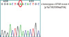
On July 30, 2010, a 25-yr-old man was admitted to our hospital because of bilateral loin pain and nausea just after severe physical activity, and that was 2nd episode since 1 month before. He had played football and jogging on a regular base since he was 20 yr old, but there was no problem. He had completed military service in Korea without any medical problem. Five days prior to his visit, he had run thousands of meters for an employment test of a local security company. Just after the test, he got vomiting and loin pain. He endured the symptoms for a few days, but oliguria developed the day before he was admitted. He had experienced same symptoms after same physical activity test, 1 month ago. On admission, he had oliguria. Height and weight were 171 cm and 81 kg. Blood pressure was 143/85 mmHg and body temperature was 36.3℃. Physical examination did not reveal any abnormalities except bilateral costovertebral angle tenderness. Laboratory tests showed the following: hemoglobin 12.6 g/dL, hematocrit 35.9%, leukocyte count 6,160/µL with normal differentiation, platelet 209,000/µL, total protein 6.7 g/dL, serum sodium 139 mEq/L, potassium 4.4 mEq/L, chloride 107 mEq/L, BUN/Cr 29.0/4.31 mg/dL, CK/LDH 87/259 U/L, CRP 1.24 mg/dL, uric acid 2.0 mg/dL, spot urine sodium 45 mEq/L, urine myoglobin(-), 24-hr urine sodium 179 mM/day, potassium 3 mM/day, chloride 231 mM/day, uric acid 517 mg/day, Cr 1.68 g/day, fractional excretion of sodium 4.6%, and fractional excretion of uric acid 66%. The kidney sonographic image showed left kidney/right kidney 12.3/12.9 cm and no abnormal echo texture. After 9 days of admission, marked hypouricemia became apparent with the improvement of renal function: BUN was 18.4 mg/dL, serum creatinine 1.66 mg/dL, and serum uric acid 1.4 mg/dL. Gene analysis was done under the diagnosis of exercise-induced ARF associated with idiopathic renal hypouricemia. Peripheral blood sample for gene analysis was obtained from the patient. DNA sequence analysis of the exon 1, 3, and 4 (hot spot for mutations) of the URAT1 gene was undertaken. Gene analysis revealed a homozygous nonsense mutation (c.G774A, p.Trp258Stop) in the exon 4 of the URAT1 gene (Fig. 1). After 11 days of admission, he was discharged with the improvement of renal function (BUN 17.1 mg/dL, serum creatinine 1.14 mg/dL).
DISCUSSION
Uric acid is the end product of the metabolism of purine compounds. Contrary to the vast majority of mammalian species, the human homolog of the mammalian uricase gene is structurally modified to an unexpressed (pseudogene) state. As a result, normal humans have serum urate concentrations approaching the theoretical limit of solubility of urate in serum (6.8 mg/dL). With the exception of minor non-specific contributions from peroxidases and catalases, human tissues do not have the ability to metabolize urate. Thus, in order to maintain homeostasis, urate must be eliminated by the gut and the kidney. Hypouricemia is defined as serum uric acid level below 2 mg/dL. This can be the result from the condition which uric acid production decreases. But more commonly, it results from increased renal uric acid excretion (2). The causes of increased urate clearance include medications with uricosuric properties, total parenteral hyperalimentation, and defects in renal tubular transport such as Fanconi syndrome. Some cases of familial hypouricemia result from a loss-of-function mutation in SLC22A12, the gene that encodes for URAT1.
The pathogenesis and clinical details of ARF associated with renal hypouricemia remain unknown. In 2002, Ishikawa (1) reported two types of exercise-induced ARF: one is the well-known myoglobin-induced ARF, and the other is new type of ARF with severe loin pain which develops after anaerobic exercise (ALPE). ALPE develops after anaerobic exercise such as 200-meter tracking racing. The patient in this case who ran hundred meters without rest also developed ALPE. The relationship between hypouricemia and ALPE is not fully understood. However, recently a urate/organic anion exchanger (URAT1) has been identified and characterized (7, 8). The URAT1 is a highly urate-specific and distinct organic anion exchanger and is encoded by SLC22A12, a gene residing on chromosome 11q13. A defect in the SLC22A12 gene is the known major cause of idiopathic renal hypouricemia. Most patients with idiopathic renal hypouricemia have loss-of-function mutations in SLC22A12. Although many different mutations have been reported in SLC22A12, W258X and R90H are the common mutations in Korea (9-11). In our case, 258th amino acid substituted guanine to adenine, which means stop codon resulting from a homozygous nonsense mutation (c.G774A, p.W258X) in the exon 4 of the URAT1 gene (Fig. 1).
Uric acid is a powerful antioxidant, and is a scavenger of oxygen free radicals (12) which has been suggested to injure nephron segments, especially proximal tubules. In patients with renal hypouricemia, the uric acid pool is very small, the static intracellular concentration of uric acid is low, and the total amount of uric acid mobilized into proximal tubular cell is also very small, although the daily urinary excretion of uric acid is usually normal (13). During exercise, the production of oxygen free radicals increases, and the increase of muscular blood flow results in the decrease of renal blood flow (14, 15). This phenomenon may lead to severe vasoconstriction in patients with hypouricemia, which is likely to occur when the intracellular concentration of uric acid is low (13). In addition, oxygen free radicals may be over-produced after the recovery of renal blood flow in patients with severe vasoconstriction compared to those in healthy people, as shown in ischemia-reperfusion models (16, 17). For these reasons, patients with renal hypouricemia may be prone to develop ARF. There was a seasonal/monthly variation in the occurrence of ARF according to 54 renal hypouricemia induced ARF patients survey in Japan (18). In that study, ARF episodes were found to occur predominantly in May, September and October. This trend was especially marked in patients who had ARF episodes induced by short distance racing. The seasonal/monthly variation seems to coincide with the months in which most athletic meetings are held in Japan. Therefore, ARF in hypouricemic patients may be associated with specific exercise such as short-distance races. In our case, the patient had no specific symptoms after aerobic exercise such as jogging or soccer, but ARF developed after severe anaerobic exercise. This can be explained by ischemia-reperfusion models.
 XML Download