

 PDF
PDF ePub
ePub Citation
 Print
Print


INTRODUCTION
Since first described by Quain in 1847 (1), hundreds of abdominal aortic stenosis have been reported with many variations in anatomy. Though, the complete absence of infrarenal aorta, seen in presenting case, is extremely rare. Fitzpatrick reported similar case in a 15-yr-old boy (2). This represents the first case of complete segmental occlusion of infrarenal aorta in the old patient.
CASE REPORT
A 52-yr-old woman presented to the clinic with one-year history of bilateral claudication. Mild hypertension was diagnosed one year earlier, but has been well without medication. She complained right knee joint pain and frequent oral ulcers without genital ulcers. Physical examination revealed normally developed female. Superficial arterial dilatations were seen on her abdomen. There were neither murmurs nor bruits in the chest and in the abdomen. Both dorsalis pedis arterial pulses were weak. Blood pressures were measured at 130/80 mmHg in the forearms and at 100/70 mmHg in the thighs, with ankle-brachial indexes of 0.77.
Urinalysis, blood cell counts, liver function tests and serum creatinine were normal. Serologic tests including C3, C4, venereal disease research laboratory test (VDRL), and Immunoglobulin G were unremarkable except mild elevated erythrocyte sedimentation rate (ESR) of 22 mm/hr and low tittered anti-nuclear antibody of 1:80 with speckled pattern.
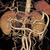
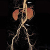
Computed tomographic angiography showed an abrupt segmental occlusion of infrarenal aorta (Fig. 1). Both renal arteries were intact. The abdominal aorta was reconstituted by multiple collaterals, such as marginal artery of Drummond, the anastomosis between superior mesenteric and inferior mesenteric artery, bilateral epigastric, lumbar, and abdominal superficial arteries.
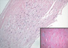
Operative findings revealed absence of infrarenal aorta replaced with fibrous band (Fig. 2). The pathology revealed fibromyxoid degeneration (Fig. 3). Polytetrafluoroethylene graft was interposed between the subdiaphragmatic thoracic aorta to the iliac bifurcation. The claudication resolved, and ankle-brachial index became 1.1. For her knee joint arthritis, she was started with methylprednisolon of 8 mg and non-steroid anti-inflammatory drug. After negative results of Pathergy test and human lymphocyte antigen B 51, we ruled out the Behcet's disease and she tapered the medications to stop successfully. She wanted the antihypertensive drug for mild hypertension and was taken amlodipine at 5 mg daily.
One year later, computer tomographic angiography showed patent bypass graft and decreased collateral vessels (Fig. 4).
DISCUSSION
The etiology of abdominal aortic stenosis is poorly understood. The young age of most of the patients suggests that the malformation may be congenital. Maycock first proposed an unequal fusion of embryonic aorta as a congenital mechanism (3). In first month of fetus, the paired primitive dorsal aortas fuse to form the abdominal aorta. Incomplete fusion due to infection or inflammation may lead of kinking of the aorta resulting to permanent constriction. Congenital rubella has been suggested for such vascular anomaly (4, 5). In Alagille syndrome which is congenital pulmonary artery stenosis transmitted in an autosomal dominant inheritance, deletion of the Jagged 1 gene has been suggested to relate with abdominal aortic coarctation (6). Acquired etiologies were also suggested in fibromuscular dysplasia or Takayasu's arthritis (7, 8). In our patients, several inflammatory presentations including the knee joint arthritis, recurrent oral ulcers, and elevation of ESR have suggested the possibility of Behcet's disease. Though, the pathologic study showed only fibromyxoid degeneration without any evidences of vasculitis. We ruled out the Behcet's disease after the negative study of Pathergy test. Considering the complete absence of infrarenal aorta without any inflammatory reaction, congenital hypoplasia thought be most proposal cause in this patient.
In review of cases for abdominal aortic stenosis, coarctation, hypoplasia, and middle aortic syndrome were used diversely. The term "coarctation" implies a congenital anomaly, and "hypoplasia" is to longer segment of coarctation. Middle aortic syndrome is the clinico-anotomical entity involving long segmental narrowing of the abdominal aorta and various visceral arteries irrespective of its etiology. We described this case by "hypoplasia", because 4 cm of infrarenal aorta was absent.
Presenting symptoms of abdominal aortic hypoplasia are related to the extent of the stenosis, and the collateral system. In case involving renal artery, patients typically present with uncontrolled hypertension. Infrarenal aortic hypoplasia can give rise to claudication when the collateral circulation is not sufficient. An abdominal aortogram via angiography, ultrasound, computed tomography, or magnetic resolution imaging can be used to diagnose and to determine the location and extent of disease. In case of renal artery stenosis, surgical intervention is aimed to treat severe hypertension and to preserve renal function. Medically treated patients have an average life expectancy of 32 yr (9). Claudication and aneurysmal dilatation also need surgical correction. In this case, as the subrenal aortic stump was too short, graft was interposed between the subdiaphragmatic thoracic aorta to the iliac bifurcation. Her hypertension seems not related to the aortic disease.
Claudication is not uncommon. Careful physical examination for inspection of dilated superficial arteries and auscultation of bruits can provide a clue for rare causes of ileofemoral insufficiency such as abdominal aortic stenosis. When suspected, an angiography is mandatory to diagnose, and surgical intervention is necessary to improve severe claudication.

 XML Download