

 PDF
PDF ePub
ePub Citation
 Print
Print


INTRODUCTION
Human epidermis is a keratinized and stratified squamous epithelium, forming an impermeable physical barrier. It not only protects the host against invading pathogens, but also provides an effective barrier to noxious external stimuli and dehydration (1, 2). To accomplish these functions, keratinocyte progenitor cells in the basal layer proliferate, move upward, and then differentiate into cornified cells. As the keratinocytes differentiation process takes place along a pathway that leads to cell cycle arrest and terminal differentiation, a complex program of gene expressions should be coordinated (3, 4).
To gain insight into the molecular events involved in keratinocytes differentiation, investigators have adopted several experimental techniques. For examples, hundreds of known and novel proteins have been identified in epidermis and cultured keratinocytes using two-dimensional polyacrylamide gel electrophoresis (5). Also thousands of genes were identified by serial analysis of gene expression (SAGE) (6, 7). Although SAGE allows quantitative as well as qualitative analysis of a large number of genes, the tag sequences provided by SAGE are sometimes too short to match one specific gene in sequence databases. In contrast, random sequencing of a cDNA library provides long sequences that are enough to distinguish the genes originated from the same ancestors. However, it has a redundancy problem that makes it difficult to identify all the expressed genes. It is therefore advantageous to apply a normalization procedure and bring the frequency of each clone in a cDNA library within a narrow range (8). A normalized cDNA library can increase the efficiency of subtractive hybridization procedures, and can significantly enhance genomic research pursuing chromosomal assignment of expressed sequences (exon mapping) (9).
To investigate the molecular events involved in keratinocytes differentiation, we chose calcium, the best characterized factor as a differentiating agent for keratinocytes. In epidermis, it takes about 14 days for keratinocytes to undergo terminal differentiation and become dead corneocytes. Although there has been no direct evidence that chronological time progress in vitro exactly matches that of in vivo condition, we speculated that similar enough cellular events would occur in in vitro condition. Based on this assumption, in this study, we constructed a normalized library using the mRNA isolated from keratinocyte treated with calcium for 14 days and analyzed the gene expression profile.
MATERIALS AND METHODS
Skin samples
All skin samples were obtained under the informed consent of donors, in accordance with the ethical committee approval process of Chungnam National University Hospital, Daejeon, Korea. The study was conducted according to the Declaration of Helsinki principles.
Cell culture
Specimens were briefly sterilized in 70% ethanol, minced, and then treated with dispase overnight at 4℃. The epidermis was separated and placed in a solution containing 0.05% trypsin and 0.02% ethylenediaminetetraacetic acid (Gibco BRL, Rockville, MD, USA) for 15 min at 37℃. After vigorous pipetting, cells were pelleted and resuspended in a keratinocyte-serum free medium (K-SFM) supplemented with bovine pituitary extract and recombinant human epidermal growth factor (Gibco BRL). Third passaged keratinocytes (60-80% confluency) were used in this experiment. To induce differentiation, keratinocytes were incubated with K-SFM containing 1.2 mM calcium chloride.
Construction of a normalized cDNA library
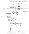
Messenger RNA was isolated using an Oligotex mRNA kit (Qiagen, Hilden, Germany). One µg of mRNA was annealed with NotI-tag-(dT)18 primer, then reverse transcribed using Superscript Reverse Transcriptase (Invitrogen, Carlsbad, CA, USA). Double-strand cDNAs were size-selected by gel filtration, followed by ligation with an EcoRI adaptor (Amersham, Buckinghamshire, UK). After digestion with NotI and EcoRI, the cDNAs were directionally cloned into the pT7T3-Pac vector and transformed into DH5αF' using electroporation. Double-strand plasmid DNAs were converted into single-strand DNAs by superinfection with helper phage M13KO7. Single-strand library DNAs were used as a template for PCR amplification with T3 and T7 primers. PCR-amplified cDNA fragments were used as the driver cDNAs in consequent subtraction procedure. For subtraction, cDNA fragments were purified, melted, and hybridized with single-strand library DNA in the presence of blocking oligonucleotides. The remaining single-strand DNAs were purified by HAP chromatography, converted into double-strand plasmids, then electroporated into bacteria to generate a normalized library (Fig. 1) (10). For identification of cloned genes, about 10,000 colonies were randomly picked, and plasmid DNAs were prepared using the Wizard Miniprep DNA purification system (Promega, Madison, WI, USA). Sequencing was performed using previously described procedures (11).
Functional categorization and reverse transcription-polymerase chain reaction (RT-PCR)
Functional categorization of the genes was performed using the Database for Annotation, Visualization and Integrated Discovery (DAVID, http://apps1.niaid.nih.gov/david/). Two µg of total RNAs were reverse transcribed with M-MLV reverse transcriptase (Promega). Aliquots of RT mixture were subjected to PCR cycles with specific primer sets. All primers used in this study were designed using Primer 3 (http://frodo.wi.mit.edu/cgi-bin/primer3/primer3_www.cgi), and verified by the BLAST to ensure that each sequence was unique to the specific target gene. The primers used in the study are as follows: MGC3200, 5'-CGACTTGAGGAAGCCCAGTA and 5'-TCATGGCTCCTCGTAGCTCT; FLJ25124, 5'-GGCTGCAAGAAGAAAACCAG and 5'-TGGTTCTTCTGGGAATCCTG; FLJ11280, 5'-AGAGCAGTTCCTGGAGACCA and 5'-GAGAACCAGAAACCCACCAA; MGC2747, 5'-GCGACCAACAGGCTAAAGAG and 5'-CCCAGAGTCGGCTGAAGTAG; FLJ22329, 5'-GATGAATGTGTGCGACAACC and 5'-TCTGGAGGTTCTGGGAAATG; FLJ20850, 5'-ATGCTAACTCGCCCTCAGAA and 5'-TTTGACCCTGTCCTTGGAAC; FLJ36445, 5'-GCCCTCACCTGACTTTGTGT and 5'-AGAGGGTGTTGACCATCTCG.; cyclophilin, 5'-CTCCTTTGAGCTGTTTGCAG and 5'-CACCACATGCTTGCCATCCA; loricrin, 5'-GTGGGAGCGTCAAGTACTCC and 5'-AGAGTAGCCGCAGACAGAGC; involucrin, 5'-CAAAGAACCTGGAGCAGGAG and 5'-CAGGGCTGGTTGAATGTCTT.
RESULTS
Gene expression profile and functional categorization
Keratinocytes cultured in a low calcium condition (less than 0.03 mM) do not differentiate, and show the similar morphology to the basal epidermal cells. When the calcium concentration is raised above 0.1 mM, these cells begin to acquire the morphological and biochemical characteristics of suprabasal cells and express several differentiation-related genes (12, 13). To better understand the molecular events in the keratinocytes differentiation process, we constructed a normalized cDNA library using the mRNA isolated from normal human epidermal keratinocytes cultured under a 1.2 mM calcium condition for 14 days. After sequencing about 10,000 clones, 4,104 independent genes were obtained, indicating that the normalization of cDNA library was successful. They consisted of 3,699 annotated genes (90.1%) and 405 expressed sequence tags (ESTs) (9.9%). Several genes implicated in keratinocytes differentiation, such as keratin 1, transglutaminase 1, and loricrin, were identified from our normalized library. In addition, many genes that had not previously been associated with keratinocytes were also isolated.
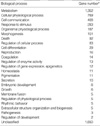
To categorize the genes in terms of biological process, we performed gene ontology analysis using the DAVID program. The functional categorization is summarized in Table 1 (a detailed gene list is available when requested). Majority of genes were classified into the categories of metabolism, cellular physiological process, and cell communication, revealing that keratinocytes differentiation is a dynamic biological process rather than a simple prerequisite for preparing the programmed cell death. Interestingly, 29 genes were classified into the cell differentiation category (Table 2), in which several genes implicated in keratinocytes differentiation, such as Notch 1 and four and a half LIM domains 1 (FHL1), were included (14, 15).
Determination of chromosomal location
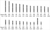
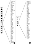
To determine the chromosomal location of the genes obtained from the normalized library, we used the Ensembl Genome Browser (http://www.ensembl.org/index.html) provided by EMBL-EBI and Sanger Institute. Of note, many of genes were located at chromosomes 1 (11.20%) and chromosome 19 (8.23%) (Fig. 2), reflecting the possibility of linkage to the epidermal differentiation complex (EDC) on chromosome 1q21 and another putative EDC on 19q13.1 (16, 17). In an attempt to identify novel keratinocytes differentiation-related genes, we focused on EST genes located on chromosomes 1q21 and 19q13.1. We found 3 ESTs on chromosome 1q21 and 4 ESTs on chromosome 19q13.1 (Fig. 3). To confirm the expression of these ESTs, we performed RT-PCR analysis using the total RNAs isolated from cultured keratinocytes treated with 1.2 mM calcium. We chose 4 time points that reflect the specific situation of differentiating keratinocytes; day 1 for the cells leaving from basal layer, day 3 for the early spinous layer, day 7 for the middle spinous layer, and day 14 for the late spinous or granular layer (4). As shown in Fig. 4, most of the ESTs were confirmed to be expressed in keratinocytes. However, they did not show differential expression during the keratinocyte differentiation process, while the well-known differentiation markers such as loricrin and involucrin increased by calcium treatment. One EST, FLJ36445, showed increased expression in a time-dependent manner (Fig. 4), suggesting that this EST is probably a novel candidate gene relevant to keratinocytes differentiation.
DISCUSSION
With the improvement and development of new techniques, such as SAGE, two-dimensional polyacrylamide gel analysis and cDNA microarray, considerable amount of knowledge about gene expression profile in keratinocytes differentiation has been obtained (6, 7, 18, 19). In this study, we further extended gene expression profile in keratinocytes treated with 1.2 mM calcium, using normalized cDNA library.
As an initial step to uncover the biological process underlying keratinocytes differentiation, we previously concentrated our efforts on finding the differentially expressed genes. Adopted molecular techniques included suppression subtractive hybridization (SSH) and cDNA microarray (4, 20, 21). Although these techniques give very useful information, they have limitation in screening the entire expressed genes, as there are many genes that do not show significant changes at the mRNA level. Also, because there may be a significant number of important genes modulating cellular activity with small copies of mRNA (for example, 1-15 copies per cell), it is necessary to adopt more specific technology for profiling the whole expressed genes. Normalized cDNA library is very useful method for this purpose, because it provides information on the expressed genes in certain condition, regardless of their transcriptional changes and/or the amount (copy number) per cell. In this study, we adopted a normalization technique and tried to profile as many expressed genes as possible in keratinocytes. We just sequenced about 10,000 clones in this study, and identified 4,104 non-redundant genes. The expression of many genes identified in this study have never been studied yet in keratinocytes, thus our results provide clues on which to base further investigations of the molecular function in keratinocytes. Since analysis of the human genome sequence has identified approximately 25,000-30,000 protein-coding genes (22), it is estimated that we can analyze all the expressed genes in keratinocytes if we sequence ten times more clones from our normalized library. Although the 4,104 non-redundant genes obtained here are not sufficient to profile all the expressed genes, they provide important information with regard to the keratinocytes differentiation. For example, chromosomal allocation reveals that many genes expressed in keratinocytes cultured under a 1.2 mM calcium condition are from chromosome 1 and 19. An interesting point is that chromosome 19 is relatively small in size as compared with other chromosomes, nonetheless, it is second only to chromosome 1 in terms of number of genes that are expressed in keratinocytes cultured in a high calcium concentration. As previously shown, chromosome 1q21 harbors epidermal differentiation complex (EDC), in which many gene families involved in terminal differentiation of keratinocytes are located (16, 23, 24). In addition, another probable EDC is suggested to be located at chromosome 19q13.1, in which the genes encoding keratinocyte differentiation-associated protein (Kdap), suprabasin, and dermokine-α/-β, are mapped (17, 25-27). Therefore, our results reflect somehow the relationship between the amount of genes and the EDCs on chromosome 1q21 and 19q13.1.
Actually, at the beginning, we wanted to compare the gene expression profile between non-differentiated and fully-differentiated keratinocytes using the normalized libraries. To this end, we chose two time points; those are 0 day (non-treated) and 14 day (calcium-treated). We also made a normalized library using the mRNA isolated from calcium non-treated undifferentiated keratinocytes. We just sequenced about 5,000 clones and acquired about 2,000 independent genes. In terms of identifying the differentially expressed genes, actually, the normalized cDNA library is not an efficient method as compared with subtraction technologies, because that it needs tremendous amount of sequencing to cover whole expressed genes. Although it is inadequate to compare the whole gene expression profiles, nevertheless, our preliminary analysis showed certain difference. As mentioned before, in our study, the number of genes identified from chromosome 19 is second to that of chromosome 1. To compare the gene numbers expressed between undifferentiating and calcium-treated keratinocytes, we counted the genes isolated from a normalized library that was made using the mRNA isolated from calcium-non-treated undifferentiated keratinocytes. The results showed that clear difference in percentage of expressed genes from chromosome 19 between undifferentiated and calcium-treated keratinocytes (4.8% vs. 8.2%). Furthermore, one EST located on chromosome 19q13.1, FLJ36445, showed differential expression during keratinocytes differentiation (Fig. 4), strengthening the notion that there may be two or more EDCs in the genome. The identifying new EDCs will be an important further study.
In summary, we made the normalized library and examined the gene expression profile. Our results provide additional information on the expressed genes during keratinocyte differentiation, which offers considerable clues for future studies.

 XML Download