

 PDF
PDF ePub
ePub Citation
 Print
Print


INTRODUCTION
Despite the improvements in surgical techniques, immunosuppression, and antimicrobial agents, infection remains one of the important complications after solid organ transplantation (1-3). The incidence of infectious complications is especially high in patients who undergo liver tansplantation (LT), because patients requiring this procedure tend to be in poorer condition and the combination of the surgical procedure and the use of immunosuppression after LT is associated with a higher risk of infection than after transplantation of other solid organs.
Children who undergo organ transplantation have a higher incidence of viral infection and a poorer outcome than adults, due to the high incidence of primary infection, especially with cytomegalovirus (CMV) (4) and Epstein-Barr virus (EBV) (5), in younger seronegative patients. Therefore studies of infection after LT in children differ markedly from studies in adults. LT in children more frequently involves living than cadaveric donor tissue, because of the scarcity of size-matched deceased donors for small children. However, studies of infectious complications after living donor liver transplantation (LDLT) in children are relatively rare (6-9).
Although LDLT was initially developed to overcome the shortage of donor livers for children (10), it has now been adapted for adults in areas with donor shortages (11). Ideally LDLT is associated with the improvement of graft and patient survival because the condition of these patients is better, due to shorter waiting time, use of elective surgery, and better graft viability due to short ischemic time (12).
Studies of infection following pediatric LDLT are necessary to improve management after LT and to provide better patient outcomes. We therefore investigated infectious complications after pediatric LDLT.
MATERIALS AND METHODS
Patients
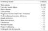
We enrolled 95 children (38 boys, 57 girls) who underwent LDLT at the Asan Medical Center from 1994 to 2004. Median age of the patients at LT was 22 months (range, 6 months to 15 yr). Sixty children underwent LDLT for biliary atresia, 15 for acute liver failure, 7 for Wilson disease, 3 for unexplained liver cirrhosis, 2 for progressive familial intrahepatic cholestasis, 2 for ornithine transcarbamylase deficiency, and 6 for other causes (Table 1).
Perioperative management
The left lateral segment or left lobe grafts of the liver were implanted into most of the children according to routine procedures (13). Those patients were managed in the intensive care unit for 5 to 14 days postoperatively, including isolation, and were usually discharged from the hospital without complications 3 to 4 weeks after surgery. For 2 days prior to surgery, all patients underwent selective bowel decontamination with oral neomycin, erythromycin, and mycostatin. All patients received intravenous prophylactic antibiotics with Unasyn® (ampicillin plus sulbactam, 150 mg/kg/day) and cefotaxime (100 mg/kg/day) before surgery and for 5 days postoperatively. All patients received CMV prophylaxis with intravenous ganciclovir (6 mg/kg/day) for 14 days after surgery, followed by oral acyclovir (40 mg/kg/day) for 6 months. All patients also received mycostatin (500,000 U/day) for antifungal prophylaxis and sulfamethoxazole-trimethoprim (8 mL/kg/day for 3 days) for Pneumocystis jiroveci prophylaxis for 6 months after surgery.
Immunosuppression
Maintenance immunosuppression regimens consisted of a triple cyclosporine-based drug regimen, which included azathioprine and prednisolone (PD) for the first 11 patients; all other patients received a dual tacrolimus-based drug regimen that included PD. Cyclosporine was administered intravenously, and then orally, with the dosages adjusted to maintain trough level at 250-350 ng/mL for the first 6 months, 100-250 ng/mL for the next 6 months and 50-100 ng/mL thereafter. Oral tacrolimus was administered, with the dosages adjusted to maintain trough level at 10-15 ng/mL for the first 2 weeks, 5-10 ng/mL for the next 2 months, and 5 ng/mL thereafter. Methyl PD was administered at a dosage of 10 mg/kg intraoperatively, and tapered to 0.3 mg/kg on day 7. Beginning on day 8, all patients were administered oral PD, starting at a dosage of 0.3 mg/kg/day. In patients given the cyclosporine regimen, PD was changed into on alternate days indefinitely and in patients given the tacrolimus regimen, PD was tapered during the next 3-6 months for patients. All patients given the cyclosporine regimen were also administered azathioprine for 3 months, at an average dosage of 1 mg/kg/day. Children who experienced acute rejection were given methylprednisolone pulse therapy; if there was no response, daclizumab, azathioprine, and mycophenolate mofetil were administered according to response.
Diagnosis of infection
Bacterial and fungal infections were diagnosed based on their clinical manifestations and the isolation of organisms. Viral infections were diagnosed based on clinical manifestations, serology, the shell vial antigen method for CMV, immunohistochemical staining for CMV, EBV-encoded RNA in situ hybridization, and, since 2003, quantitative polymerasechain reaction for CMV and EBV load monitoring. Primary infection was diagnosed when seroconversion occurred, from negative preoperatively to positive postoperatively. Reactivation was diagnosed when there was a four-fold increase in titer or anti-IgM antibody was detected. Hepatitis B virus infection was not analyzed in this study.
RESULTS
Overall incidence of infections
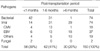
We observed 150 infections in 67 of the 95 (70%) patients (1.57 infections per patient); 74 infections in 43 patients were bacterial, 2 in 2 patients were fungal, and 66 in 42 patients were viral. Most of the bacterial and fungal infections occurred within 1 month after LDLT, whereas most viral infections occurred after 1 month (Table 2).
Bacterial infections
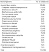
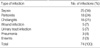
We observed 74 episodes of bacterial infection in 43 children, 42 (57%) within 1 month after LDLT and 73 (99%) within 6 months. Of the 74 infections, 41 (55%) were with Gram negative organisms, 32 (44%) were with Gram positive organisms, and 1 (1%) was with an anaerobic organism. The most common bacterial pathogens were Klebsiella pneumoniae (16%), Pseudomonas aeruginosa (16%), coagulase-negative Staphylococcus (15%), and Enterobacter faecium (10%) (Table 3). The most common clinical manifestations of bacterial infection were septicemia (33%), peritonitis (25%), and cholangitis (21%) (Table 4).
Fungal and parasitic infections
Only two episodes of fungal infection, in 2 patients, were documented. Both patients had a history of vancomycin treatment and both organisms were isolated from ascites. No episodes of parasitic infection were observed.
Viral infections
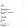
Seventy four episodes of viral infection occurred in 42 patients, including 37 episodes of EBV infection, 18 of CMV, 10 of rotavirus, 2 of respiratory syncytial virus (RSV), 2 of enterovirus, 2 of rubella virus, 1 of influenza virus, 1 of herpes simplex virus and one of parainfluenza virus (Table 5). Of the 18 episodes of CMV infection, 14 were primary infections and 4 were reactivations. Of the involved patients, 9 had hepatitis, 2 had gastroenteritis, 2 had pneumonia, and 7 were asymptomatic. Most of the infection occurred between one to 6 months. Thirty seven patients experienced EBV infection; 11 were primary infections and 26 were reactivations. Of these patients, 9 had posttransplant lymphoproliferative disease (PTLD), 21 had hepatitis, 4 had gastroenteritis, 3 had lymph node involvement, 2 had infectious mononucleosis with fever, and 9 were asymptomatic. Eight of the 9 cases of PTLD developed within 24 months, and 5 of these 9 patients died of PTLD. PTLD did not occur in any of the 18 patients who underwent LDLT in the last 2 yr, following routine EBV viral load monitoring. RSV infection occurred in 2 patients; one occurred immediately after LDLT and this patient died of acute respiratory distress syndrome.
DISCUSSION
Studies of infectious complications after pediatric LT are not common, although one study (14) reported that the incidence of infection after pediatric LT was 1.36 episodes per patient, a result similar to our finding of 1.57 episodes per patient. In addition, a recent study that included 2,291 children (1) reported that severe infectious complications occurred in 52% of patients within 15 months after LT.
Most infections occurring within 1 month after LT have been found to be bacterial (7, 9, 15, 16). Bacterial infections occurring soon after LT have not been associated with increased mortality (17). Patients undergoing transplantation of other solid organs, using similar immunosuppressive regimens, showed similar patterns of infectious complications relative to the interval after transplantation (9). While most infections during the first month after transplantation are associated with perioperative problems, those occurring 1 to 6 months after transplantation are associated with the use of immunosuppressive drugs (18). Most CMV infections occur between 3 and 8 weeks after transplantation (3, 4, 19), whereas most EBV infections occur within 6 months. Infections are relatively uncommon after 6 months and those that occur are primarily associated with chronic rejection, retransplantation, or large doses of immunosuppressive therapy. We observed similar findings after pediatric LDLT, in that 57% of bacterial infections occurred within 1 month and PTLD associated with EBV occurred more frequently after 6 months.
We found a similar incidence of infection with Gram positive and Gram negative organisms, whereas others (14) have reported that the incidence of infection with Gram positive bacteria was much higher (78%). We found that the most common Gram positive infection was with coagulase negative Staphylococcus, whereas the most common Gram negative bacteria were enteric bacteria such as Klebsiella pneumoniae, Enterobacter cloacae, Proteus mirabilis, E. coli, and Citrobacter freundii, findings similar to those of other studies (15, 18). Enteric Gram negative bacteria frequently cause infections in patients who undergo transplantation of abdominal organs, because these bacteria can cause peritonitis by spillage from the intestinal lumen into the peritoneum, as well as causing ascending cholangitis by reflux from the intestinal lumen into the bile duct during surgery (3, 20-22). Therefore preoperative selective bowel decontamination and careful attention during surgery are necessary to decrease the rate of infection. Candida albicans was the only source of fungal infection in our study, occurring in only two patients. While fungal infection usually occurs during the third week after transplantation (8), both cases of fungal infection observed here occurred earlier, around 10 days after LDLT. However, both were associated with prolonged preoperative antibiotic usage for prophylaxis of bacterial infection.
Bacterial and viral infection is an important cause of mortality after LT (1), with CMV reported to be the main source of viral infection (14). We also found that the infection was the main cause of mortality after LDLT, with 50% of the deaths we observed associated with infection. All of these deaths were associated with viral infection; none was directly related to bacterial infection. Only one patient died of CMV infection, which may have been associated with the relatively lower incidence of seronegative recipients (18 of 88 recipients, 20%, unpublished data). Of the other deaths, 5 were from PTLD and one was from RSV infection.
Early detection of EBV infection by quantitative EBV load monitoring and subsequent reduction in the use of immunosuppressive agents has been reported to reduce the development of PTLD and mortality (23). We found that the incidence of PTLD decreased dramatically after adoption of EBV load monitoring. However, our institution has used the latter routinely only for 2 yr. Further study, for a longer period of time, is needed.
In conclusion, this study showed that infectious complications after pediatric LDLT were similar to those observed after pediatric cadaver donor liver transplantation. While bacterial infection occurs mainly during the immediate postoperative period and does not affect patient mortality, viral infection occurs mainly after 1 month and is a main cause of patient death after pediatric LDLT. Appropriate attention should be paid to reduce the morbidity and mortality from infectious complication according to the period of time after pediatric LDLT.
 XML Download