Abstract
We had three cases of Moraxella osloensis meningitis. The species identification was impossible by conventional and commercial phenotypic tests. However, we could identify the species using the 16S rRNA gene sequencing. Determination of clinical significance was difficult in one patient. All three patients recovered by appropriate antimicrobial therapy.
Moraxella osloensis, an oxidase-positive, nonmotile, asaccharolytic, aerobic gram-negative coccobacilli is a part of the normal flora of the human respiratory tract (1, 2). The organism is difficult to identify because of presence of several other species with similar phenotypic characteristics. Species of some glucose non-fermenting gram-negative bacilli are not only difficult to identify by routine methods, but also the clinical significance is not always easy to interpret. Clinical significance of M. osloensis isolates even from normally sterile site may be difficult to determine, because the organism rarely cause invasive infections. We report three cases of M. osloensis meningitis in two children with no known underlying disease and in one adult cancer patient.
A 4-yr-old girl was admitted to the Pediatric Department on May 27, 2002, with a 1-day history of headache, fever (38.1℃), abdominal pain, and vomiting (Table 1). Her past medical history was unremarkable and she was up-to-date with her immunizations. Her physical and neurological examinations were unremarkable.
Laboratory tests revealed a peripheral white blood cell (WBC) count of 19,800/µL (91% neutrophils), a hemoglobin level of 11.6 g/dL, and a platelet count of 356,000/µL. Cerebrospinal fluid (CSF) had a hazy appearance with an increased WBC count of 890/µL (99% neutrophils), but no significant change in glucose concentration of 78 mg/dL, and a protein level of 31 mg/dL (Table 2). Routine blood chemistry results were within the reference range. A chest radiograph was normal.
The patient was treated with cefotaxime and ampicillin for 1 week. The patient became afebrile after 2 days of treatment, and her other symptoms also improved. The CSF analysis showed a decreased WBC count to 9/µL from 890/µL at hospital day 3. She was discharged on 9th hospital day without complications.
A 15-yr-old boy was admitted to the Neurology Department on May 18, 2005, with a 3-day history of headache and 2 days of generalized petechiae, nausea, and vomiting (Table 1). His past medical history was unremarkable. On admission, his oral temperature was 36.9℃, heart rate was 93 beats/min, and blood pressure was 125/82 mmHg. He was alert and complained about neck stiffness. There were no motor or sensory deficits, and the rest of his physical examination was unremarkable.
A laboratory investigation at the time of admission revealed a peripheral WBC count of 30,290/µL (96% neutrophils), a hemoglobin level of 15.4 g/dL, and a platelet count of 151,000/µL. CSF had a cloudy appearance with an increased protein concentration of 575 mg/dL, and a WBC count of 35,500/µL (90% neutrophils) and a decreased glucose concentration of 10 mg/dL (Table 2). Routine blood chemistry results were within the reference range. A chest radiograph was normal.
The patient was treated with ceftazidime and netilmicin for 2 weeks. His clinical condition was improved after 3 days, and the CSF analysis showed a decreased WBC count and protein concentration to 4,150/µL and 90 mg/dL from 35,500/µL and 575 mg/dL, respectively. On 13th hospital day, the CSF WBC count decreased to 130/µL, and the patient was discharged without complications on 18th hospital day.
An 81-yr-old man with a known history of pancreatic cancer (March, 2004) and liver cirrhosis due to hepatitis C virus infection (June, 2004) was admitted to the Emergency Department on June 6, 2005, with drowsy mental status for one day. On admission, he had icteric sclera (Table 1).
Laboratory investigation revealed a peripheral WBC count of 6,950/µL (73% neutrophils), a hemoglobin level of 12.8 g/dL, and a platelet count of 60,000/µL. CSF was clear with a protein level of 38 mg/dL, a slightly increased WBC count of 9/µL, and a glucose concentration of 76 mg/dL (Table 2). Routine blood chemistry showed increased blood urea nitrogen/creatinine of 41.8/2.2 mg/dL, ammonia of 287 µg/dL and total bilirubin of 3.1 mg/dL. The other test results were unremarkable.
The patient was treated with cefotaxime for 1 week. On hospital day 3, the CSF WBC count decreased to 0/µL. He was managed conservatively and was discharged 18th hospital day without complications.
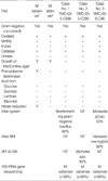
In case no. 2 many gram-negative coccobacilli were detected on smear of a CSF. In cases no. 1 and no. 3, bacteria were not detected on smears. The culture of CSF specimens from case no. 2 on blood and chocolate agar plates yielded heavy growth of medium sized (0.5-1 mm) grayish colonies after overnight CO2 incubation. In case No. 1, moderate growth was observed after overnight CO2 incubation. In case no. 3, several colonies were detected after 2 days incubation. The Gram-stained smear of colonies revealed gram-negative coccobacilli in all three cases. The isolates were oxidase positive and did not oxidize glucose. The correct species identification was not achieved not only by the conventional biochemical tests, but also by the API ID 32 GN system (profile 00000-000002), and Vitek2 GN system (bioMerieux, Marcy l'Etoile, France), but suggested Moraxella spp. 16S rRNA gene sequencing (3) identified all three isolates as M. osloensis (≥98% agreement with GenBank accession no. AF005190) (Table 3). The antimicrobial susceptibility test performed by the disk diffusion method (4) showed all three isolates were susceptible to ampicillin, cephalothin, cefoxitin, cefotaxime, ceftazidime, cefepime, levofloxacin, imipenem, amikacin, netilmicin, and gentamicin.
Members of the genus Moraxella are oxidase-positive, nonmotile, and asaccharolytic coccobacilli. Genus Moraxella consists of seven species, M. atlantae, M. canis, M. catarrhalis, M. lacunata, M. lincolnii, M. nonliquefaciens, and M. osloensis (1). Graham et al. (5) reported that M. osloensis was common and the most frequent isolate from blood and CSF among clinical isolates of Moraxella spp. It has been rarely reported to cause local or invasive infections, including osteomyelitis, meningitis, bacteremia, pneumonia, and catheter associated infection (6-11).
A PubMed search yielded four published cases of M. osloensis meningitis. Two patients had known predisposing factors for infection such as cerebrospinal fluid shunt and C8 complement deficiency, but the remaining two patients had no known underlying disease (12). The children in case 1 and 2 had no underlying diseases. These children had typical meningitis symptoms such as headache, neck stiffness, nausea, and vomiting, an elevated WBC count, and increased protein in CSF analysis. In case No. 1, we could not totally exclude the coexistence of viral and bacterial meningitis because we did not test virus culture nor virus antigen assay. The adult patient (case No. 3) had underlying pancreatic cancer and liver cirrhosis. As this patient had drowsy mental status on admission, CSF examination and culture were performed to rule out meningitis. A possible explanation of low CSF WBC count was due to lower peripheral WBC count resulting from various cytotoxic therapies. The clinical diagnosis of this patient was hepatic encephalopathy because of high blood ammonia and high total bilirubin level. This patient was also diagnosed as bacterial meningitis because of M. osloensis growth from CSF. With appropriate antimicrobial agents' treatment, peripheral blood WBC count and CSF WBC count decreased.
Identification of an unusual bacterial isolate is a very difficult problem in a clinical microbiology laboratory. Moraxella spp. grows well in capnophilic conditions, but grows slowly in ambient air which may lead to delays in diagnosis. Commercially available identification kit systems are sometimes unable to reliably identify unusual microorganisms. Using conventional biochemical tests and two commercial identification kits, we were able to infer the genus Moraxella. However, we could not definitively identify the species because the results of various biochemical tests are similar among the seven Moraxella spp.
Only with 16S rRNA gene sequence analysis, we were able to definitively identify the isolates as M. osloensis (3). This new method consists of PCR amplification and sequencing of the 16S rRNA gene in bacteria, followed by a comparison of the 16S rRNA gene sequence with those registered in GenBank. The case no. 1 and no. 3 isolates matched with more than 99% and the case no. 2 isolate matched with more than 98% agreement with M. osloensis.
M. osloensis is usually susceptible to penicillin G, cephalosporins, and aminoglycosides (5, 13). There are a few reports that treatment with penicillin G and ampicillin for M. osloensis bacteremia and meningitis has been successful. All three strains were susceptible to majority of antimicrobial agents, including ampicillin, cephalothin, cefotaxime, amikacin, levofloxacin, and imipenem. All patients were treated successfully with third-generation cephalosporins alone or in combination with netilmicin.
In conclusion, although M. osloensis meningitis is rare it may cause severe CNS infection in children and in adult cancer patient. We were able to definitely identify the species of the isolates only by using 16S rRNA gene sequencing. All three patients recovered by appropriate antimicrobial therapy
References
1. Schreckenberger PC, Daneshvar MI, Weyant RS, Hollis DG. Murray PR, Baron EJ, Jorgensen JH, Pfaller MA, editors. Acinetobacter, Achromobacter, Chryseobacterium, Moraxella, and other nonfermentative gram-negative rods. Manual of Clinical Microbiology. 2003. Washington: American Society for Microbiology;754–757.
2. Weyant RS, Moss CW, Weaver RE, Hollis DG, Jordan JG, Cook EC, Daneshvar MI. Identification of unusual pathogenic gram-negative aerobic and facultatively anaerobic bacteria. 1995. Baltimore: Williams & Wilkins;390–405.
3. Clarridge JE 3rd. Impact of 16S rRNA gene sequence analysis for identification of bacteria on clinical microbiology and infectious diseases. Clin Microbiol Rev. 2004. 17:840–862.

4. NCCLS. Document M100-S12. Performance Standards for Antimicrobial Susceptibility Testing; Twelfth Informational Supplement. 2002. Wayne, Pa: NCCLS.
5. Graham DR, Band JD, Thornsberry C, Hollis DG, Weaver RE. Infections caused by Moraxella, Moraxella urethralis, Moraxella-like groups M-5 and M-6, and Kingella kingae in the United States, 1953-1980. Rev Infect Dis. 1990. 12:423–431.

6. Sugarman B, Clarridge J. Osteomyelitis caused by Moraxella osloensis. J Clin Microbiol. 1982. 15:1148–1149.

7. Berger U, Kreissel M. Menigitis due to Moraxella osloensis. Infection. 1974. 2:166–168.
8. Fritsche D, Karte H, del Solar E. Moraxella osloensis as pathogen in septicemia. Infection. 1976. 4:53–54.
9. Vuori-Holopainen E, Salo E, Saxen H, Vaara M, Tarkka E, Peltola H. Clinical "pneumococcal pneumonia" due to Moraxella osloensis: case report and a review. Scand J Infect Dis. 2001. 33:625–627.
10. Han XY, Tarrand JJ. Moraxella osloensis blood and catheter infections during anticancer chemotherapy: clinical and microbiologic studies of 10 cases. Am J Clin Pathol. 2004. 121:581–587.
11. Buchman AL, Pickett MJ, Mann L, Ament ME. Central venous catheter infection caused by Moraxella osloensis in a patient receiving home parenteral nutrition. Diagn Microbiol Infect Dis. 1993. 17:163–166.

12. Shah SS, Ruth A, Coffin SE. Infection due to Moraxella osloensis: case report and review of the literature. Clin Infect Dis. 2000. 30:179–181.

13. Rosenthal SL, Freundlich LF, Gilardi GL, Clodomar FY. In vitro antibiotic sensitivity of Moraxella species. Chemotherapy. 1978. 24:360–363.


 PDF
PDF ePub
ePub Citation
 Print
Print


 XML Download