

 PDF
PDF ePub
ePub Citation
 Print
Print


INTRODUCTION
Acute thermal injuries to the esophagus are uncommon causes of esophageal disease. A candy-cane appearance is an endoscopic feature of thermal injury to the esophagus from hot liquid (1, 2). However, the clinical course of such a finding from solid food is not well documented. We treated a patient that presented with a 2-day history of odynophagia and foreign body sensation after an acute thermal injury of the esophagus from solid food (prawn). Here we present the clinical course and the endoscopic findings.
CASE REPORT
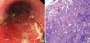
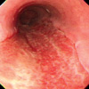
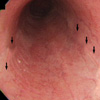
A 53-yr-old man was admitted to the hospital with a two-day history of odynophagia and a foreign body sensation. He had no medical history, and previously denied any esophageal symptoms such as dysphagia, odynophagia, or chest discomfort. Two days before admission, the patient began to experience odynophagia and a foreign body sensation in the chest after swallowing several extremely hot pieces of prawn in haste. On arrival, the vital signs were normal and laboratory tests were unremarkable. Endoscopy revealed a huge longitudinal ulcer, typical of friable hyperemic mucosa with necrotic debris with a tendency for easy to touch bleeding along the full length of the esophagus (Fig. 1A) in the posterolateral region. In addition, there was a foul odor. Biopsy specimens from the esophagus revealed marked ulceration with activated endothelial cells (Fig. 1B). The patient was treated with a parenteral nutrition regimen and intravenous pantoprazole to prevent injury from gastroesophageal reflux. The symptoms gradually improved over eight days after the initial event. At eight days after the initial event, the repeated endoscopy revealed hyperemic mucosa and an intervening whitish pseudomembrane (Fig. 2). The patient was started on liquids and progressed to a soft diet, and was then discharged on oral pantoprazole for 1 months. Eight weeks after the event, endoscopy showed normal esophageal mucosa with the rim of the previous ulcer scar (Fig. 3).
DISCUSSION
Here we present the clinical course of serial endoscopy of an acute thermal injury of the esophagus caused by extremely hot solid food. This is a rare cause of esophageal disease (1-6). There is only one prior similar report with the endoscopic and histological characteristics of an injured esophagus due to hot liquids (3). However, there is no same case due to solid foods reported in the medical literature. Although candy cane esophagus with alternating pink and white linear bands of esophageal mucosa is well known feature of esophageal thermal injury, most of the prior caustic agents were hot liquids. In our case, the typical candy cane appearance was not observed during the clinical course. Most early endoscopic findings of the initial injury during the first week showed a predominant whitish pseudomembrane (3). However, our findings, 2 days after the initial event, showed hyperemic areas with marginal necrotic debris, which may have resulted from rupture of bullae. We assume that the appearance of the candy cane esophagus was due to the flow of hot liquid whereas that of burn or ulcer was due to the pressure effect of hot solid. Therefore the different causes of thermal injury might make the different endoscopic findings and the endoscopic image of a candy cane appearance may not be characteristic of an acute esophageal thermal injury due to hot solid foods. There are very few data on the management strategies for acute esophageal thermal injury. Although most reported series are case reports, conservative management with anti antisecretory treatment such as a proton pump inhibitor (PPI) or histamine2-receptor antagonist (H2RA) to prevent further injuries from gastric acid was underwent without severe complications (3-6). Most cases were treated for 1 months except for only one case for 14 days (6).
In conclusion, with the history of hot food ingestion, esophageal symptoms, a foul odor on examination, and deep linear ulcers on the posterior aspect of the esophagus the diagnosis of an acute thermal injury of esophagus should be considered.
 XML Download