

 PDF
PDF ePub
ePub Citation
 Print
Print


INTRODUCTION
When performing an electrodiagnostic evaluation of the upper extremities, it is important to know the components of the brachial plexus (BP) and the knowledge regarding the myotomes or peripheral innervations of muscles sampled by needle electromyography (EMG) is essential for the accurate diagnosis of focal nerve injury and cervical radiculopathy. The ulnar nerve is simply the continuation of the medial cord into the upper limb, and the C8 and T1 root levels are the primary constituents of the ulnar nerve (1-3). However, many clinicians believe the C7 root to be present in the ulnar nerve, and abnormal findings could be observed in muscles innervated by the ulnar nerve, especially the flexor carpi ulnaris (FCU), in C7 radiculopathy (4-7). The anatomical background of the presence of the C7 component in the ulnar nerve is attributable to an anatomical pathway that allows neural fibers from C7 to enter the ulnar nerve by way of the so called 'lateral root of the ulnar nerve (LUN)'; however, this pathway has been mostly ignored in the standard description of BP. The prevalence is highly variable, and it has been reported at rates of 15% to 92% (5). This factor raised the need for the investigation to clarify the components of the ulnar nerve and its clinical significance. The FCU is commonly examined during needle EMG for the diagnosis of lower brachial plexopathy, ulnar neuropathy and cervical radiculopathies. However, disagreement on the myotomes corresponding to the FCU muscle may lead to an erroneous interpretation, especially in the diagnosis of patients with cervical radiculopathies.
We designed this study to verify the variations of the BP for the formation of the ulnar nerve through cadaveric dissection and to analyze the myotomes of the FCU using electrophysiologic study.
MATERIALS AND METHODS
To determine the prevalence of anatomical variations, dissection was performed on 38 arms from 19 adult cadavers (male 8, female 11). The dissection fields were confined to the infraclavicular portion of the BP. We investigated the presence of connecting branches from the lateral cord to the ulnar nerve and to the medial cord.
We also reviewed the results of electrodiagnostic studies performed at the Electrodiagnostic Laboratory of the Department of Physical Medicine and Rehabilitation, Korea University Anam Hospital between January 2006 and May 2008 and collected electrodiagnostic data in patients diagnosed with cervical radiculopathy. Cervical radiculopathy was diagnosed by needle EMG when abnormal spontaneous activities and/or long-duration, large amplitude, polyphasic motor unit action potentials (MUAPs) were present in at least three muscles of different peripheral nerve innervations but of the same myotome. Only patients with radiculopathy located in a single cervical root segment, C6, C7, or C8, were included in this study. Patients diagnosed with cervical radiculopathy concomitant with brachial plexopathy, focal neuropathy, or polyneuropathy were excluded. There were 244 cases of single-level radiculopathy at the C6, C7, or C8 segment, and we selected cases that fulfilled the following electrodiagnostic criteria: 1) inclusion of the FCU on needle EMG, 2) abnormal needle EMG findings in at least 3 muscles of each myotome other than the FCU in each level of cervical radiculopathy, 3) abnormal spontaneous activities and/or large amplitude motor unit potentials with a reduced recruitment pattern of at least 1 muscle other than the FCU in each level of radiculopathy.
A total of 106 cases were included in the final analysis, and the patients were divided into three groups of C6, C7, or C8 radiculopathy. We investigated the proportion of neuropathic needle EMG findings of FCU ('neuropathic FCU') in patients with C6, C7, or C8 radiculopathy. To avoid the inclusion of equivocal cases, the criteria for neuropathic findings were defined as the presence of abnormal spontaneous activities at rest and/or large amplitude MUAPs, as well as reduced recruitment patterns during voluntary contraction.
RESULTS
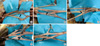
Branches from the lateral cord to the medial cord or ulnar nerve were observed in 5 (13.1%) of the 38 arms examined in the cadaver study. The connection was from the lateral cord to the ulnar nerve in 4 cases and from the lateral cord to the medial cord in one case (Fig. 1). All of the cases were unilateral.
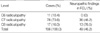
Electrophysiological analyses of 106 cases of single-level radiculopathy revealed that the numbers of patients with C6, C7, or C8 radiculopathy were 11 (10.4%), 78 (73.6%) and 17 (16.0%), respectively. In contrast to the relatively low incidence (13.1%) of C7 contribution to the ulnar nerve in the cadaveric study, electrodiagnostic data demonstrated that the incidence of neuropathic EMG findings of FCU was high in patients with C7 radiculopathy, as it occurred in 36 (46.2%) of 78 cases. An 'neuropathic FCU' was observed in 13 (76.5%) of 17 cases of C8 radiculopathy, but it was not detected in any case of C6 radiculopathy (Table 1).
DISCUSSION
The brachial plexus innervates the entire upper limb and most of the muscles in the shoulder girdle. Focal lesions of the BP typically produce unique patterns of electrodiagnostic findings. The gross anatomy of the BP is most complex, and its internal topography is very intricate. Though major anomalies of the BP are relatively rare, Kerr (8) described 29 types of variations of the BP. The most commonly reported anomalies involve a shift of the root origins of the BP fibers by one root in the rostral or caudal direction, pre-fixed plexus (C4-C8), post-fixed plexus (C6-T2), or expansion (C4-T1) (3).
Regarding LUN, some literatures depicted the LUN in the figure of the BP (5, 9), but the presence of the LUN and its clinical significance were not emphasized. The incidence of LUN is highly variable (9, 10). Bowden et al. (10) reported that the incidence of C7 to the medial cord was 12.5% (14/168) while the incidence of C7 to the ulnar nerve was 3.0% (5/168). However, Fuss (9) reported that the incidence of LUN as 56% in their anatomical study. In our study, the incidence of lateral cord to the ulnar nerve was 10.5% (4/38), the incidence of lateral cord to the medial cord was 2.6% (1/38), and the total incidence was 13.1% (5/38). Our results are very similar to those reported by Bowden et al. (10), though a connecting branch to the ulnar nerve was more common than to the medial cord in our study. Though some authors have suggested that the starting point of the terminal nerve, which lies 3 cm beyond the cord or transition site at which the terminal nerves exit the axilla, there are no anatomic dividing lines between the cord and the terminal nerve of the BP, which makes it difficult to differentiate the proximal portion of the terminal nerves from the distal cords (11). Regarding the clinical significance of LUN, Wilbourn (12) denied the clinical impact of various anomalies because the incidence is questionable and the anomaly generally does not affect the segmental innervations of the branches of the BP. On the other hand, Dumitru and Zwarts (5) argued that LUN is not an anomaly, but rather a major portion of the BP that is ignored in the "standard" description.
Radiculopathy is one of the common reasons for referral to the electrodiagnostic laboratory. The most commonly affected nerve root in the cervical region is C7, which accounts for approximately 31-81% of all cervical radiculopathies (5, 13, 14). Electrodiagnosis of radiculopathies primarily relies on the finding of membrane instability in a myotomal distribution on needle EMG. Basic knowledge on segmental innervation of the muscle is fundamental for the accurate diagnosis of radiculopathies, but there is some disagreement among the myotomes because of the way they were derived, anatomic dissection, clinical examination or EMG findings (2, 7, 14). These discrepancies regarding the myotomes can lead to confusion in the electrodiagnosis of the exact level of cervical radiculopathy. The medial cord is composed of the C8 and T1 sensory and motor fibers, but when the lateral cord or C7 root sends fibers to the ulnar nerve, C7 radiculopathy can produce abnormalities in muscles innervated by the ulnar nerve, such as the FCU. The FCU is frequently used in the diagnosis of C8 radiculopathy, but less commonly in the diagnosis of C7 radiculopathy (14). Though there are insufficient data regarding the prevalence of FCU involvement in C7 radiculopathy, FCU demonstrated neuropathic findings in about 28% of electrodiagnostically confirmed cervical radiculopathies (15) and in 34-65% of C7 radiculopathy (14). In our study, 46.2% of the patients with C7 radiculopathy demonstrated neuropathic findings in the FCU. These results are 3 times higher than the predicted value of 13.1% based on our anatomical dissection. This discrepancy could result from both anatomical and electrodiagnostic factors. From an anatomical aspect, the contribution of the C7 root to the ulnar nerve could be achieved through another anatomical variation, such as prefixed BP. The prevalence of pre-fixed BP is also highly variable, and it ranges from 2% to 48% (16, 17). If the prefixed BP exists, the medial cord is formed mainly by the C7 and C8 roots, and the C7 component in the FCU muscle could be high. Another anatomical factor to be considered is that the field of dissection in this study was confined to the infraclavicular plexus in order to detect anomalies of cords and terminal nerves. Thus, abnormalities of the supraclavicular plexus could not be encountered in our study. Electrodiagnostic factors are investigated by the criteria for 'neuropathic' findings in the interpretation of needle EMG. The prevalence of abnormal EMG findings can be affected by the criteria whether it is restrictive or inclusive. We used relatively restrictive criteria, abnormal spontaneous activities and/or large amplitude MUAPs with reduced recruitment, for the interpretation of abnormal needle EMG findings in order to avoid disagreement. If we had included only the patients showing abnormal spontaneous activity, the number of cases showing abnormal findings in the FCU might have decreased. One study (15) reported that the prevalence of spontaneous activity in at least one muscle in cervical radiculopathy is 88%, and it decreased to 65% in 2 or more muscles. They reported that spontaneous activities in the FCU muscle were less common and only observed in 16% of subjects with electrodiagnostically confirmed cervical radiculopathies. Therefore, inclusion of the spontaneous activities as a common denominator is not feasible.
In this study we attempted to elucidate the clinical significance of LUN through anatomical studies and electrophysiological analysis of myotomes corresponding to the FCU. This result suggested that LUN is not an uncommon finding. In addition, the C7 root commonly contributed to myotomes of the FCU in nearly one-half of our cases, but there was no C6 component in the FCU. This information would be helpful for the diagnosis of C7 radiculopathy and some types of brachial plexopathies involving the middle trunk or lateral cord. In the electrodiagnosis of suspected cervical radiculopathies, the presence of abnormal needle EMG findings in the FCU combined with other C7 myotomal muscles could be diagnosed as C7 radiculopathy.
Based on the results of this study, LUN is not an uncommon variant, and it is a conceivable pathway for the C7 root to the ulnar nerve. Although it was not emphasized previously in clinical anatomy, LUN is an important variation for electromyographers. The C7 myotome in the FCU is relatively common, and needle EMG of this muscle may provide more information for the electrodiagnosis of cervical radiculopathy and brachial plexopathy.
 XML Download