

 PDF
PDF ePub
ePub Citation
 Print
Print


INTRODUCTION
Green tea, a world wide consuming beverage, is known to have the beneficial effects on the cardiovascular system. In fact, the intake of green tea in human is proposed to be associated with a lower incidence of coronary artery disease (1) and myocardial infarction (2). In addition, there is evidence that the consumption of green tea reduces mortality due to cardiovascular disease in a large population prospective study (3). Furthermore, polyphenol (-)-epigallocatechin gallate (EGCG), the most prominent catechin of green tea, appears to attenuate myocardial ischemia/reperfusion (I/R) injury in animal models (4, 5). However, there is scanty information available about the molecular mechanisms involved in the EGCG-induced cardioprotection so far.
Meanwhile, the intracellular calcium ([Ca2+]i) overloading has been implicated as a primary event in cell injury and necrosis in myocardial I/R injury (6). Opening of adenosine triphosphate-sensitive potassium (KATP) channels prevents the [Ca2+]i overload induced by myocardial I/R injury (7). Recent studies have suggested that mitochondrial KATP (mKATP) channels are responsible for the marked cardioprotective effect of myocardial ischemic preconditioning and pre-ischemic drug treatment to reduce infarct size (8, 9). Furthermore, there is accumulating evidence that the cardioprotective effects of KATP channel openers are associated with mKATP activation (10).
In this regard, it's reasonable to hypothesize that the activation of mKATP channels may participate in the cardioprotective effect by EGCG. Therefore, the purpose of this study was to determine the involvement of mKATP channels in EGCG-induced pharmacological preconditioning in isolated rat hearts.
MATERIALS AND METHODS
The experimental procedures and protocols used in this study were reviewed and approved by our institutional animal care and use committee of Keimyung Universiry Dongsan Medical Center.
Chemicals and antibodies
Pentobarbital sodium (Entobar®) was purchased from Hanlim Pharmacy (Yongin, Korea). EGCG and a selective blocker of mKATP channels (5-hydroxydecanoate, HD) were purchased from Sigma-Aldrich Chemical (St. Louis, MO, USA). A nonselective KATP channel blocker glibenclamide (GLI) was obtained from Tocris Bioscience (Ellisville, MO, USA). 2,3,5-Triphenyltetrazolium chloride (TTC) was also obtained from Sigma-Aldrich Chemical. Fluorescent polymer microspheres were purchased from Duke Scientific Corp. (Palo Alto, CA, USA). Other chemicals were obtained from Sigma-Aldrich Chemical.
Langendorff isolated heart perfusion preparation
Male Wistar rats, weighing 280-330 g obtained from Korea Takonic (Seongnam, Korea), were used throughout the experiments. They received 100 mg/kg of pentobarbital sodium and 300 IU of heparin intraperitoneally. Hearts were isolated and perfused with modified Krebs-Henseleit solution containing (in mM) 118.5 NaCl, 4.7 KCl, 1.2 MgSO4, 1.8 CaCl2, 24.8 NaHCO3, 1.2 KH2PO4, and 10 glucose, as described previously (11). In order to induce regional ischemia, the proximal portion of left coronary artery (LCA) was first localized between the left atrial appendage and the right ventricular outflow tract. This was then followed by the passage of a 6-0 polypropylene suture around the major trunk of the LCA or its prominent branches. The ends of the thread were passed through a small piece of PE50 tube to form a snare. All hearts were then allowed to stabilize for at least 30 min. Ischemia was induced by pulling the snare and then fixing it by clamping the tubing with a small hemostat and was confirmed by regional cyanosis, a substantial decrease in left ventricular developed pressure (LVDP), or a fall in coronary flow. Reperfusion was initiated by releasing the snare. Hearts experiencing ventricular fibrillation (VF) usually revert spontaneously to sinus rhythm. VF lasting more than 45 sec was treated with finger flick cardioversion until a perfusing rhythm was obtained. No pharmacological agents were used for defibrillation.
Assessment of cardiac function
The functional recovery by EGCG was compared with control hearts. To do this, an air-bubble free, Krebs-Henseleit buffer-filled elastic balloon made of polyethylene plastic connected to a pressure transducer with tubing was inserted into the left ventricle (LV) through the left atrial appendage. The balloon was coupled to a graded threaded micro-syringe and balloon volume was adjusted to give a left ventricular end-diastolic pressure (LVEDP) of 5-10 mmHg at the beginning of the experiment. LVDP was calculated as the difference between the left ventricular systolic pressure (LVSP) and LVEDP. Coronary flow was measured by a timed collection of the perfusate dripping from the right heart into a graduated cylinder. Cardiodynamic data, including heart rate, LVSP, LVEDP, and the maximum and minimum of first derivative of left ventricular pressure (+dP/dtmax and -dP/dtmin) were continuously recorded with the MP150 pressure transducer (BioPac Systems, Santa Barbara, CA, USA), and analyzed using analysis software (Acqknowledge, version 3.9.0.). The rate-pressure product (RPP) was calculated as LVDP×heart rate (12).
Experimental protocol
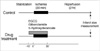
All hearts were subjected to 30 min of regional ischemia and 2 hr of reperfusion and each group consisted of at least 7 hearts. Isolated hearts were assigned randomly to one of the following groups; 1) Control, no other intervention either before or after LCA occlusion; 2) EGCG1, 1 µM of EGCG; and 3) EGCG10, 10 µM of EGCG (Fig. 1). Following subgroup hearts were used to determine the involvement of KATP channels in EGCG induced cardioprotetion using a nonselective KATP channel blocker GLI and a selective mKATP channel blocker HD; 4) EGCG+GLI EGCG with 10 µM GLI; 5) EGCG+HD; EGCG with 100 µM HD; 6) GLI alone; and 7) HD alone. Compounds were infused for 40 min, from 10 min before to the end of index ischemia. Drugs were diluted with Krebs-Henseleit solution to the required final concentrations, as described above, on the day of each experiment. The concentrations of KATP channel blockers used in this study were based on previous studies for isolated working rat hearts (13, 14).
Exclusion criteria
We decided prospectively that any hearts with a heart rate less than 250 beats/min, or coronary flow more than 18 mL/min or less than 8 mL/min at the end of stabilization would be excluded from the study. Hearts failing to develop LVSP of more than 80 mmHg when the LVEDP was kept 5-10 mmHg and hearts exhibiting arrhythmia during stabilization period also discarded.
Determination of area at risk and infarct size
At the end of each experiment (2 hr after reperfusion), the area at risk (AAR) and area at necrosis (AN) were measured as described in our previous study (11). In brief, LCA perfusion circuit was reoccluded, and diluted fluorescent polymer microspheres with 2-9 µm in diameter were infused to demarcate the AAR. The hearts were weighed, frozen at -20℃ for 1-3 hr, and cut into 2 mm thick transverse slices using a rat heart slice matrix (Zivic Instruments, Pittsburgh, PA, USA). The slices were incubated in 1% TTC in sodium phosphate buffer (pH 7.4) at 37℃ for 20 min. The slices were immersed in 10% formalin to enhance the contrast between viable (stained) and necrotic (unstained) tissue. LV was removed from the remaining tissue and then the LV slices were compressed to a uniform 2 mm thickness by placing them (basal side) between two glass plates separated by a 2 mm space. The AAR of myocardium was identified by illuminating the slices with U.V. light as the tissue without fluorescence. The AN (unstained by TTC) and AAR (no fluorescence) zone regions were traced on a clear acetate transparent sheet and quantified with the Image Tool (UTHSCSA Image Tool, version 3.0). The areas were converted into volumes by multiplying the areas by slice thickness. The volume of AN was expressed as a percentage of the AAR volume. All measurements were performed in a blinded fashion.
Statistical analysis
Data are expressed as means±SEM. Data analysis was performed with a personal computer statistical software package (SPSS for Windows, Release 12.0; SPSS Inc, Chicago, IL, USA). One-way analysis of variance with Tukey's HSD post hoc test was used to determine whether any significant differences existed in cardiodynamic parameters and infarct measurements between groups. Chi square analysis was used to test differences in the incidence of VF. Differences were considered to be statistically significant when P values were less than 0.05.
RESULTS
A total of 53 rat hearts were used for this experiment. All hearts presented in results were perfused within 30-40 sec after excision. Three hearts were excluded from data analysis for the following reasons: coronary flow >18 mL/min (n=1), LVDP <80 mmHg (n=1), or heart rate <250 beats/min (n=1) during the stabilization period. Therefore, we report the data for 50 successfully completed infarct experiments.
Incidence of ventricular fibrillation
No heart had VF during ischemia or after 30 min of reperfusion. VF occurred during early reperfusion and appeared either as a transient self-limiting episode or as a persistent episode which need finger flick cardioversion. VF occurred in 17 of 50 hearts (3/8 in control. 2/7 in EGCG1, 2/7 in EGCG10, 2/7 in EGCG+GLI, 2/7 in GLI, 3/7 in EGCG+HD, and 3/7 in HD) during early reperfusion. There was no significant difference in the incidence of VF among groups.
Determination of infarct size
Table 1 presents body weight, heart weight, and LV volume. There were no significant differences in these parameters among the groups. Table 1 also presents risk volume data for the groups. Risk volume averaged 0.275 cm3 to 0.320 cm3 and was not statistically different among groups (P>0.05). The mean AAR for each experimental group ranged from 53.6% to 63.2% for LV volume. There were no significant differences in the ratio of AAR to LV volume among the groups and these results indicates that all groups were subjected to equivalent degrees of regional ischemia. This result means that the changes in the infarct size observed between various groups in our experiments were not related to the degree of ischemic area.
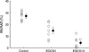
As shown in Fig. 2, infarct volume in the control hearts was 27.2±1.4% of the AAR zone. The ratio of infarct volume/ischemic volume (AN/AAR) in our control hearts was smaller than those reported by others (11, 12), however, this is in agreement with our recently reported study (13).
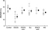
EGCG treatment during ischemic period significantly reduced myocardial infarction (14.5±2.5% in 1µM EGCG and 4.0±1.7% in 10 µM EGCG, P<0.001 vs. control). The AN/AAR was more markedly attenuated by 10 µM EGCG than by 1 µM EGCG (P=0.003). To confirm the involvement of KATP channels in infarct limitation effect by EGCG, we examined whether a nonselective KATP channel blocker GLI and a selective mKATP channel blocker HD could limit the anti-infarct effect by 1 µM EGCG. The infarct limitation effect by 1 µM of EGCG was totally abrogated by 10 µM GLI (24.6±1.5%, P<0.001 vs. EGCG). Similarly, 100 µM HD also aborted the anti-infarct effect of EGCG (24.1±1.2%, P<0.001 vs. EGCG) (Fig. 3). Treatment of control hearts with GLI or HD alone had infarct size of 23.7±2.5% and 21.0±1.0%, respectively, which were not different compared with the infarct size in the untreated control hearts (P>0.05).
Cardiac function recovery
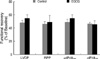
The baseline coronary flow and cardiac function parameters are given in Table 2. There were no significant differences in coronary flow and hemodynamic indexes concerning heart rate, LVDP, RPP, +dP/dtmax, and -dP/dtmin at baseline among the groups. There were no significant differences in coronary flow and heart rate among groups throughout the experiments (data not shown). The LVDP, RPP, +dP/dtmax, and -dP/dtmin after 2 hr of reperfusion in control hearts were 48.0±3.3%, 46.1±3.6%, 49.1±3.8%, and 46.7±2.4%, respectively, of the baseline levels. The functional recovery parameters by EGCG after 2 hr of reperfusion were not different with control hearts (Fig. 4).
DISCUSSION
The treatment of EGCG, a major polyphenolic substance found in green tea, during ischemia significantly attenuated the infarct size after 2 hr of reperfusion in our isolated rat hearts. The current results confirm that EGCG treatment during ischemia protects against myocardial infarction. The infarct limitation effect by EGCG was totally abolished by a nonselective KATP channel blocker GLI, indicating the involvement of KATP channels in anti-infarct effect by EGCG. We further tested the hypothesis that the involvement of mKATP channels on EGCG-induced anti-infarct effect. The infarct limitation effect by EGCG was totally attenuated by a selective mKATP channels blocker HD. This result indicates, for the first time, that EGCG treatment during ischemia mediates mKATP channels, and thereby reduces infarct size in isolated rat hearts. In our isolated rat hearts, 1 µM EGCG treatment during ischemia significantly reduced the infarct volume (-50%) and the infarct limitation effect was more prominent in 10 mM EGCG treatment (-85%) compared to control hearts. In a previous study, Townsend et al. (5) firstly reported that 100 µM of EGCG perfusion prior to index ischemia lessened the extent of infarct size approximately 27% against the whole heart in the isolated rat hearts. We do not know the exact reason of the difference in the degree of anti-infarct effect between our and Townsend group's results, but the perfusion timing of the EGCG and the infarct measurement may account for it. We perfused EGCG from 10 min before to the end of index ischemia but they perfused EGCG 30 min before undergoing myocardial ischemia. Besides we expressed the infarct volume as a percentage of the AAR volume of the LV volume, but they measured the infarct size by measuring the infarct area against the whole heart including right ventricle. Otherwise, it may be attributable to the fact that high concentrations of EGCG (100 µM) directly inhibits sarcolemmal KATP (sarcKATP) channels via interacting with lipid membrane (14), thereby lessens its protective effects. It is suggested that oral ingestion more than 50 cups of green tea is required to achieve 10 µM EGCG concentration in the human plasma (15), and oral intake of green tea for 3 cups in fasting state and 10 cups in feast state can achieve 1 µM EGCG concentration in human plasma (16). In this regard, it is considered that EGCG concentration more than 10 µM may not be suitable to clinical settings to evaluate the efficacy of EGCG in myocardial ischemic injury. In our preliminary study, application to the bath solution of natural green tea extract containing a 1 µM EGCG during ischemic period significantly attenuated infarct volume of the LV volume (10.3±1.1%, n=8) compared with untreated myocardial I/R rat hearts (27.4±2.1%, n=8, P<0.05). This is the reason that we used 1 µM EGCG for further experiment to investigate the involvement of KATP channels in the infarct limitation effect induced by EGCG, although the anti-infarct effect was more prominent in 10 µM EGCG preconditioned hearts.
It is well known that [Ca2+]i overload is closely correlated with myocardial damage and cell death during ischemia (17). In myocardium, sarcKATP channels were originally postulated to participate in salvage from irreversible I/R injury, because their opening would produce an increase in the outward potassium current leading to shortening of action potential duration, which would in turn reduce the Ca2+ influx through voltage-dependent Ca2+ channels and increase the time during which the Na+/Ca2+ exchanger would operate to extrude Ca2+ from the cell (18). We have reported previously that EGCG can modulate sarcKATP channels via reducing its sensitivity to ATP, which closes the channel, and to phosphatidylinositol polyphosphates (PIP), which open the channel (19). The potency of EGCG to reduce the channel ATP sensitivity is revealed to be greater in the cardiac-type channels than in the beta cell-type channels. Therefore, it can be speculated that reduced ATP sensitivity of cardiac KATP channels by EGCG may be, at least partially, related with EGCG-mediated cardioprotection. However, this hypothesis should be further determined because the channel PIP sensitivity can also be reduced by EGCG, resulting in the channel be difficult to open.
It is well accepted that ischemia causes increases in intramitochondrial calcium concentrations ([Ca2+]m), resulting in further calcium-dependent events leading to cell damage (20). As calcium enters the mitochondria during ischemia, water follows, leading to mitochondrial matrix swelling and eventually mitochondrial rupture and myocyte death. The opening of mKATP channels results in mitochondrial K+ influx, expansion of the mitochondrial matrix volume, and a reduction of the inner mitochondrial membrane potential established by the proton pump. This change is expected to decrease the driving force for Ca2+ influx, therefore attenuating [Ca2+]m overload under conditions (such as I/R) in which cytosolic calcium is increased (21). Therefore, opening of mKATP channels has been proposed to play an important role in cardiac protection such as in ischemic and pharmacological preconditioning (22). The current results, indicating that the EGCG-induced anti-infarct effect is totally inhibited by HD, suggest that mKATP channel opening by EGCG during ischemia may be directly responsible for cardioprotection. Tissue salvage by an anti-infarct intervention would lead to an improvement in cardiac function. The increase in [Ca2+]i is likely to explain the depressed ability of I/R hearts to generate contractile force (23, 24). However, reduction in infarct size is not always associated with an improvement in functional recovery due to the continued stunning or the possibility that the salvaged tissue is not yet normal (25). In a previous study, preischemic infusion of EGCG (100 µM) significantly ameliorated cardiac function during postischemic phase (5). However, no significant difference was observed in the treatment of 1 µM EGCG, which is more reliable concentration in clinical settings, during ischemia compared to control group in our isolated rat hearts, although there was significant infarct limitation effect. The interaction between mitochondrial permeability transition pores (mPTP) and the mKATP channels was previously suggested by findings indicating that diazoxide (an mKATP channel opener) -induced preconditioning was inhibited by an mPTP opener atractyloside (26, 27). A role for mPTP inhibition was also suggested in pharmacological preconditioning produced by volatile anesthetics desflurane (28, 29). Therefore, the possibility of the involvement of mPTP by EGCG treatment should be determined in the future.
In conclusion, perfusion of EGCG during ischemia significantly reduces infarct size after reperfusion and the mKATP channels play a crucial role in EGCG induced cardioprotection.
 XML Download