Abstract
Clinicopathological characteristics and prognostic factors of mucinous carcinoma (MC) were compared with invasive ductal carcinoma-not otherwise specified (IDC-NOS). Clinicopathological characteristics and survivals of 104 MC patients were retrospectively reviewed and compared with those of 3,936 IDC-NOS. The median age at diagnosis was 45 yr in MC and 47 yr in IDC-NOS, respectively. The sensitivity of mammography and sonography for pure MC were 76.5% and 94.7%, respectively. MC showed favorable characteristics including less involvement of lymph node, lower stage, more expression of estrogen receptors, less HER-2 overexpression and differentiated grade, and better 10-yr disease-free survival (DFS) and overall survival (OS) (86.1% and 86.3%, respectively) than IDC-NOS (74.7% and 74.9%, respectively). Ten-year DFS of pure and mixed type was 90.2% and 68.8%, respectively. Nodal status and stage were statistically significant factors for survival. MC in Koreans showed similar features to Western populations except for a younger age of onset than in IDC-NOS. Since only pure MC showed better prognosis than IDC-NOS, it is important to differentiate mixed MC from pure MC. Middle-aged Korean women presenting breast symptoms should be examined carefully and evaluated with an appropriate diagnostic work-up because some patients present radiologically benign-like lesions.
Mucinous carcinoma (MC) of the breast is not a common disease and the incidence of MC was reported to range from 1% to 6% of all primary breast cancers (1-4). MC of the breast was characterized by a large amount of mucin production and in general, defined as having a mucinous component of 50% or more (5, 6). Those lesions had different features from invasive ductal carcinoma-not otherwise specified (IDC-NOS). MC usually occurs in postmenopausal women. Median age at diagnosis was older than 60 and older than that of IDC-NOS (4, 6). Furthermore, MC showed more favorable clinicopathological characteristics, such as lower incidence of nodal metastasis, higher expression of estrogen and progesterone receptors, and differentiated grade (2, 6, 7). Therefore, prognosis of MC is better than that of IDC-NOS with 10-yr survival rates better than 80% (1, 8).
Histologically MC is classified into 2 subgroups based on the degree of cellularity, which are pure type and mixed mucinous-ductal type (9). However, cut-off values of the mucinous component for defining pure type MC was not standardized, and later, others divided MC into type A and type B (10). The former is the classical type with large amounts of mucins and the latter is a variant type with neuroendocrine differentiation or feature of signet ring cells (5, 10).
Prognostic significance of clinicopathological characteristics in MC patients is not well established, because of its low incidence rate and lack of a standard definition. Treatment guidelines for MC are mostly extrapolated from data based on invasive ductal carcinoma (IDC) without clear validation. As incidence of breast cancer was significantly increasing in Korea (11, 12), it was necessary to investigate incidence rates, clinicopathological characteristics, and prognosis of MC to prepare an adequate treatment guideline. But there are few studies on MC and information is limited. In this study, we investigated the clinicopathological characteristics and treatment patterns for MC and compared survivals of MC with those of IDC-NOS. We evaluated the impact of those parameters on outcomes of MC.
We retrospectively reviewed the data of 4,932 breast cancer patients who were treated at the Department of Surgery, Yonsei University College of Medicine in Seoul, Korea between January 1986 and December 2006. Of them, 104 patients (2.1%) were diagnosed with MC. These patients were compared with 3,936 patients with IDC-NOS who were treated during the same period. Pathologic slides of 74 patients were available and breast pathologists reviewed their slides. However, slides of the remaining 30 patients were not available and we reviewed these cases through previous pathologic reports. Pure type MC was defined as having a mucinous component of more than 90% and specialized pathologists with extensive experience in breast pathology performed a pathologic slide review. Sixty-one (82.4%) of 74 patients were categorized as having pure type MC.
Data regarding patient demographics, histopathology of primary tumor, treatment patterns, and survivals were obtained by reviewing medical records. Patients were treated with either mastectomy or lumpectomy and axillary lymph node dissection or sentinel lymph node biopsy with local radiotherapy. After completion of surgery, adjuvant treatments were administered as indicated based on international guidelines. Tumor stage was based on the 6th American Joint Committee on Cancer (AJCC) criteria. Histological type and grading followed the World Health Organization (WHO) classification. Ten percent or more of positively stained cells was used as the cut-off for estrogen receptor (ER) and progesterone receptor (PR) positivity. HER-2/neu staining was scored from 0 to 3+ according to the guideline suggested for HercepTest™ (DAKO, Glostrup, Denmark) (13). HER-2/neu immunohistochemical staining was considered positive when strong (3+) membranous staining was observed, whereas cases with 0 to 1+ were regarded as negative. Because fluorescent in-situ hybridization test had not been performed routinely, cases with equivocal (2+) staining were excluded in statistical analyses. Clinical follow-up included history taking, physical examination, laboratory tests and radiologic imaging every 6-12 months.
The differences between the 2 groups were evaluated by chi-square test. Survival curves were determined and plotted using the Kaplan-Meier method and group differences in survival time were investigated by log-rank test. P values less than 0.05 was considered statistically significant. SPSS for Windows (version 13.0, SPSS Inc., Chicago, IL, USA) was used for all statistical analyses.
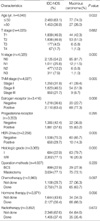
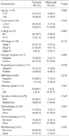
Clinicopathological characteristics and treatment patterns of all patients are summarized in Table 1. Median age at diagnosis was 47 yr in all patients (range, 20-93 yr), and age at diagnosis was much younger in MC patients than in IDC-NOS patients with statistical significance (P=0.022); 45 yr old for MC patients and 47 yr old for IDC-NOS patients. Among 74 patients whose slides were available for reviewing, 61 patients (82.4%) were pure MC and 13 (17.6%) were mixed mucinous-ductal carcinoma. Mammographic and sonographic findings according to Breast Imaging Reporting and Data System (BI-RADS) category were investigated. Mammographic and sonographic findings were available in 21 and 38 pure MC patients, respectively and 5 and 5 mixed type MC, respectively (Table 2). In pure type MC, sensitivity of mammography and sonography was 76.5% and 94.7%, respectively. However, in mixed MC, the sensitivity of mammography and sonography was 100% in both modalities.
Eighty-five (81.7%) of 104 MC patients had no axillary lymph node involvement and more MC patients had lower tumor stage than IDC-NOS patients (P=0.000 and 0.005, respectively). Expressions of ER and PR were higher in MC than in IDC-NOS; 77.3% and 63.2%, respectively, but ER only showed a statistical significance (P=0.008). Of the 70 patients whose status of HER-2/neu was available, only 10 (14.3%) showed HER-2/neu overexpression. MC showed a favorable histologic grade. Tumor size and treatments patterns including operation methods, adjuvant chemotherapy, hormonal therapy, and radiation therapy were not statistically different from IDC-NOS, but less chemotherapy and more hormone therapy were applied to MC patients due probably to more favorable histologic features associated with higher ER expression in MC. Clinicopathological characteristics and treatment patterns of pure MC patients are similar to those with the mixed type except tumor size, histologic grade, and radiation therapy (Table 3). More pure MC patients had smaller in size and differentiated histologic grade than mixed type. More radiation therapy was applied to pure MC because more pure MC patients received breast-conserving surgery.
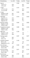
The median follow-up duration was 63.5 months in all patients (range, 0-262 months). There were 9 disease relapses and 6 deaths during follow-up in MC and 5- and 10-yr disease-free survival (DFS) rates were 93.3% and 86.1%, respectively (Fig. 1A). Five- and 10-yr overall survival (OS) rates were 93.5% and 86.3%, respectively (Fig. 1B). Ten-year DFS and OS rates of IDC-NOS were 74.7% and 74.9%, respectively. MC showed better survival outcomes than IDC-NOS with statistical significance (Fig. 1, P=0.012 for DFS and 0.005 for OS). In pure MC, the 10-yr DFS and OS rates were 90.2% and 91.3%, respectively and 10-yr DFS and OS rates of the mixed type were 68.8% and 100%, respectively. DFS and OS rates were not statistically different between pure and mixed types (Fig. 2, P=0.360 and 0.374, respectively) due probably to the small sample size. Univariate analysis for DFS and OS according to clinicopathological characteristics in MC revealed that lymph node status and tumor stage were statistically significant factors for survival (Table 4).
In stage-matched analysis for OS, MC showed a trend toward better survival than IDC-NOS (Fig. 3), but only the stage II group showed statistical significance. This was probably due to the small sample size and short follow-up duration. In age-matched analysis for OS, MC showed a better survival rate than IDC-NOS in the patient group whose age was 50 or younger (Fig. 4). Among MC patients whose pathology slide was available for reviewing, survivals of MC according to the subtype were compared with IDC-NOS (Fig. 2A, B). Pure MC showed a better DFS than IDC-NOS (P=0.037) and there was similar DFS patterns between mixed type MC and IDC-NOS (P=0.795). Pure MC showed a better OS than IDC-NOS (P=0.049) but differences of OS between the mixed type and IDC-NOS did not reach statistical significance in our study (P=0.143).
Incidence of breast cancer in Korea has significantly been increasing in recent years. It accounted for 16.8% of all female cancers in 2002 and incidence of breast cancer in Korean women is anticipated to increase (11, 14, 15). Besides incidence, the percentage of early-stage breast cancer has steadily increased (16). Screening mammography and use of ultrasonography play an important role in detection of breast cancers in earlier stages (17). Since 1996, it was recommended that all Korean women over 40 yr old underwent a mammography every 1 or 2 yr (18). From 1996 to 2004, the incidence of stage 0 and I breast cancers increased remarkably by 128.6% and 81.6%, respectively (16). However, because of the low incidence rate and small number of studies on specific type of breast cancer including MC, limited information is available regarding epidemiology, clinical, radiological and pathological features, risk factors, and prognosis. It is necessary to investigate incidence rate, clinicopathological characteristics, and prognosis of MC to prepare an adequate treatment guideline.
Most MC patients complained palpable mass in their breast, and radiologically it frequently mimics a benign lesion (19). Pure MC especially appears as a well-defined mass lesion but the mixed type shows an indistinct or microlobulated pattern (20, 21). Although image study findings were only available in a small number of patients, 23.5% of pure MC cases had false negative findings in mammography but ultrasonography showed much better sensitivity at 94.7%. Among patients available in ultrasonographic findings, most were suspicious abnormalities or highly suspicious of malignancy. These radiologically benign lesion-like characteristics of MC sometimes cause a delay in diagnosis (19). Physicians must understand the limits of screening mammography because the peak age of incidence in Korean breast cancer patients is in the 40s (16) and the sensitivity of mammography for premenopausal women is quite low (22-24). Middle-aged women presenting breast symptoms should be examined carefully and ultrasonography may be helpful.
Many Western studies reported that MC had more favorable pathological characteristics than IDC-NOS (1-4, 6, 7). They were smaller in tumor size, less axillary lymph node involvement, more expression rates of ER and PR, lower grade, lower proliferation rates and less overexpression of HER-2/neu. Another characteristic of MC was that it usually occurred in postmenopausal women (2, 4, 6), and their age at diagnosis ranged from 65 to 71 yr and older than that of IDC-NOS ranged from 59 to 61 yr (3, 6). These favorable features of MC led to better survival rates than IDC-NOS. The 10-yr OS of MC was reported at more than 80% (4, 6, 7).
In our study, the median age of MC patients was younger than IDC-NOS patients. The median age of our study population was not different from that of Korean breast cancer patients and another study reported the mean age of MC and IDC-NOS at 45.5 and 46.9 yr, respectively, which was similar to our results (12, 25). Therefore, the age of onset is quite different from that of Western MC cases. The reason for the younger age of onset in Korean women is not clear, but it might be partly due to wide use of ultrasonography, genetic or environmental factors, a cohort effect of high incidence in the younger generation or relatively easier accessibility to detect breast cancer among middle-aged women (15). However, histological features and survival rates of MC patients were similar to those of Western patients. Lymph node status was the most significant factor for survival.
MC of the breast is not a homogeneous entity and was divided into pure and mixed types (5, 10). Pure MC was associated with better survival rates than the mixed type, but survival rates of mixed type were not different from those found in IDC-NOS (2, 26, 27). Among 74 available patients with histologic subtype, there were no differences in DFS and OS between pure and mixed MC. This was probably due to the small sample size and short follow-up duration, and larger data samples with longer follow-up would be necessary to confirm these differences. Because age distribution was different between MC and IDC-NOS, we compared survival matched by age. In patients whose age was 50 yr or less, MC showed better survival than IDC-NOS (P=0.008). There was a similar trend in patient groups older than 50 yr, although it was not statistically significant (P=0.349). Some Western studies reported that age at diagnosis of MC was a significant prognostic factor (4, 6). This may be related to the older onset age in Western MC patients.
In summary, mucinous carcinoma shows more favorable histologic features and better prognosis than IDC-NOS. However, Korean MC developed at younger ages than IDC-NOS. Since only pure MC showed better prognosis than IDC-NOS, it is important to differentiate mixed MC from pure MC. Patients with mixed MC might be treated as guidelines for IDC. In pure MC patients with favorable risk factors, however, adjuvant chemotherapy might be more saved. MC clinically and radiologically mimics a benign lesion and this may lead to a delay in diagnosis. Therefore, middle-aged Korean women presenting a palpable mass should be examined carefully and the appropriate diagnostic procedures such as ultrasonography are warranted for early detection of malignant tumors.
Figures and Tables
Fig. 1
Survival curves in all patients. (A) Disease-free survival curve according to histologic type. (B) Overall survival curves according to histologic type.
IDC-NOS, Invasive ductal carcinoma-not otherwise specified.

Fig. 2
Survival curves of mucinous carcinoma patients available for histologic subtype. (A) Disease-free survival curve compared with IDC-NOS. (B) Overall survival curve compared with IDC-NOS.
MC, Mucinous carcinoma; IDC-NOS, Invasive ductal carcinoma-not otherwise specified.

Fig. 3
Stage-matched overall survival curves in all patients. (A) Stage I, (B) Stage II, (C) Stage III.
IDC-NOS, Invasive ductal carcinoma-not otherwise specified.

Fig. 4
Age-matched overall survival curves in all patients. (A) Age of 50 yr or younger, (B) Age over 50 yr.
IDC-NOS, Invasive ductal carcinoma-not otherwise specified.

ACKNOWLEDGMENTS
This work was supported by the Brain Korea 21 Project for Medical Science, Yonsei University, and in part by a grantin-aid from Novartis Korea Co., Astra Zeneca Korea Co., Dong-A Pharmaceutical Co., and Sanofi-Aventis Pharmaceutical Co.
References
1. Louwman MW, Vriezen M, van Beek MW, Nolthenius-Puylaert MC, van der Sangen MJ, Roumen RM, Kiemeney LA, Coebergh JW. Uncommon breast tumors in perspective: incidence, treatment and survival in the Netherlands. Int J Cancer. 2007. 121:127–135.

2. Andre S, Cunha F, Bernardo M, Meneses e Sousa J, Cortez F, Soares J. Mucinous carcinoma of the breast: a pathologic study of 82 cases. J Surg Oncol. 1995. 58:162–167.
3. Li CI, Uribe DJ, Daling JR. Clinical characteristics of different histologic types of breast cancer. Br J Cancer. 2005. 93:1046–1052.

4. Diab SG, Clark GM, Osborne CK, Libby A, Allred DC, Elledge RM. Tumor characteristics and clinical outcome of tubular and mucinous breast carcinomas. J Clin Oncol. 1999. 17:1442–1448.

5. Tan PH, Tse GM, Bay BH. Mucinous breast lesions: diagnostic challenges. J Clin Pathol. 2008. 61:11–19.

6. Di Saverio S, Gutierrez J, Avisar E. A retrospective review with long term follow up of 11,400 cases of pure mucinous breast carcinoma. Breast Cancer Res Treat. 2008. 111:541–547.

7. Komenaka IK, El-Tamer MB, Troxel A, Hamele-Bena D, Joseph KA, Horowitz E, Ditkoff BA, Schnabel FR. Pure mucinous carcinoma of the breast. Am J Surg. 2004. 187:528–532.

8. Page DL. Special types of invasive breast cancer, with clinical implications. Am J Surg Pathol. 2003. 27:832–835.

9. Silverberg SG, Kay S, Chitale AR, Levitt SH. Colloid carcinoma of the breast. Am J Clin Pathol. 1971. 55:355–363.

10. Capella C, Eusebi V, Mann B, Azzopardi JG. Endocrine differentiation in mucoid carcinoma of the breast. Histopathology. 1980. 4:613–630.

11. Ahn SH. Clinical characteristics of breast cancer patients in Korea in 2000. Arch Surg. 2004. 139:27–31.
12. Jung J. The Korean Breast Cancer Society. Nationwide Korean breast cancer data of 2004 using breast cancer registration program. J Breast Cancer. 2006. 9:151–161.
13. Wolff AC, Hammond ME, Schwartz JN, Hagerty KL, Allred DC, Cote RJ, Dowsett M, Fitzgibbons PL, Hanna WM, Langer A, Mc-Shane LM, Paik S, Pegram MD, Perez EA, Press MF, Rhodes A, Sturgeon C, Taube SE, Tubbs R, Vance GH, van de Vijver M, Wheeler TM, Hayes DF. American Society of Clinical Oncology. College of American Pathologists. American Society of Clinical Oncology/College of American Pathologists guideline recommendations for human epidermal growth factor 2 testing in breast cancer. J Clin Oncol. 2007. 25:118–145.
14. Shin HR, Jung KW, Won YJ, Park JG. 2002 Annual report of the Korea Central Cancer Registry: based on registered data from 139 hospitals. Cancer Res Treat. 2004. 36:103–114.

15. Son BH, Kwak BS, Kim JK, Kim HJ, Hong SJ, Lee JS, Hwang UK, Yoon HS, Ahn SH. Changing patterns in the clinical characteristics of Korean patients with breast cancer during the last 15 years. Arch Surg. 2006. 141:155–160.

16. Ahn SH, Yoo KY. Chronological changes of clinical characteristics in 31,115 new breast cancer patients among Koreans during 1996-2004. Breast Cancer Res Treat. 2006. 99:209–214.

17. Smith RA, Cokkinides V, Brawley OW. Cancer screening in the United States, 2008: a review of current American Cancer Society guidelines and cancer screening issues. CA Cancer J Clin. 2008. 58:161–179.

18. Lee SY, Jeong SH, Kim J, Jung SH, Song KB, Nam CM. Scheduling mammography screening for the early detection of breast cancer in Korean women. J Med Screen. 2007. 14:205–209.

19. Dhillon R, Depree P, Metcalf C, Wylie E. Screen-detected mucinous breast carcinoma: potential for delayed diagnosis. Clin Radiol. 2006. 61:423–430.

20. Memis A, Ozdemir N, Parildar M, Ustun EE, Erhan Y. Mucinous (colloid) breast cancer: mammographic and US features with histologic correlation. Eur J Radiol. 2000. 35:39–43.

21. Matsuda M, Yoshimoto M, Iwase T, Takahasi K, Kasumi F, Akiyama F, Sakamoto G. Mammographic and clinicopathological features of mucinous carcinoma of the breast. Breast Cancer. 2000. 7:65–70.

22. Margolese R. Screening mammography in young women: a different perspective. Lancet. 1996. 347:881–882.

23. Houssami N, Irwig L, Simpson JM, McKessar M, Noakes J. Sydney Breast Imaging Accuracy Study: comparative sensitivity and specificity of mammography and sonography in young women with symptoms. AJR Am J Roentgenol. 2003. 180:935–940.
24. Foxcroft LM, Evans EB, Porter AJ. The diagnosis of breast cancer in women younger than 40. Breast. 2004. 13:297–306.

25. Kim HJ, Kim SW, Chung KW, Chang MC, Youn YK, Oh SK, Choe KJ, Noh DY. Clinicopathologic characteristics of mucinous carcinoma of the breast. J Korean Surg Soc. 2001. 61:266–272.
26. Scopsi L, Andreola S, Pilotti S, Bufalino R, Baldini MT, Testori A, Rilke F. Mucinous carcinoma of the breast. A clinicopathologic, histochemical, and immunocytochemical study with special reference to neuroendocrine differentiation. Am J Surg Pathol. 1994. 18:702–711.
27. Komaki K, Sakamoto G, Sugano H, Morimoto T, Monden Y. Mucinous carcinoma of the breast in Japan. A prognostic analysis based on morphologic features. Cancer. 1988. 61:989–996.


 PDF
PDF ePub
ePub Citation
 Print
Print


 XML Download