

 PDF
PDF ePub
ePub Citation
 Print
Print


INTRODUCTION
Torus hyperplasia is a very rare condition also known as focal pyloric hypertrophy (1). This lesion is caused by a circular muscle hypertrophy affecting the lesser curvature near the pylorus. Pathogenesis of the lesion is unclear, some considering it as a persistence of infantile pyloric hypertrophy to adulthood, others noting that it develops within a few weeks in adult (1, 2). Some have speculated that the lesion results from chronic gastritis or from repeated spastic contractions of the area secondary to visceral-visceral reflexes that could be established as a result of local irritation stimulating afferent fibers and starting a reflex arc involving efferents that locally innervate the circular pyloric musculature (1, 3). In summary, focal hypertrophic stenosis of the pyloric antrum in adult is classified as primary or secondary types; 1) primary type being an idiopathic torus hyperplasia uncertain of origin, and 2) secondary type from a result of muscular thickening due to inflammation, ulceration, or carcinoma (4).
After the first report on 1946, torus hyperplasia has been reported as a rare disease (5). Although there are some typical findings (i.e., recognizable radiologically by narrowing and enlongation of the pyloric cancal and endoscopically by appearances resembling those of the cervix so called characteristic pyloric "cervix sign") in this benign disease (6, 7), it is difficult to confirm without resection. Moreover, it is hard to predict the prevalence of the disease because of its' rarity (3). Herein, we report a 56-yr-old man who was diagnosed as torus hyperplasia with appendiceal mucocele after visiting our hospital because of abdominal discomfort. To the best of our knowledge, this is the first case of torus hyperplasia with appendiceal mucocele which is found incidentally.
CASE REPORT
A 56-yr-old man with a history of hypertension presented with abdominal discomfort for one month. He reported no bowel habit change, nausea, vomiting, or fever, but complained of anal pain. He had a family history since his father died of advanced gastric cancer. On arrival, his blood pressure was 128/87 mmHg, heart rate 80 beats/min, respiratory rate was 20/min, and body temperature was 36.0℃. The physical examination revealed no abnormalities except rectal examination. His abdomen was soft and non-tender with normal bowel sound. On rectal examination, a fissure was noticed on 6 o'clock direction.
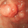
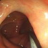
There was no remarkable finding on laboratory examinations on arrival. Upper gastrointestinal endoscopy showed a 1.0 cm sized irregular submucosal lesion proximal to the pylorus to the distal antrum on the lesser curvature. Central erosion was noticed on the surface of the lesion due to previous biopsy (Fig. 1). Cushion sign revealed negative finding, and biopsy revealed no significant abnormality. On colonoscopy examination which was performed on the same day with upper gastrointestinal endoscopy, a 1.5 cm sized protruding mass was noticed on the appendiceal orifice. This lesion revealed positive tent sign suggesting a cystic change (Fig. 2).
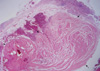
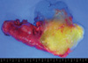
Gastrectomy and cecectomy were underwent after abdominal computed tomography. Histological section revealed marked hypertrophy of the distal circular pyloric musculature without an evidence of neoplasia (Fig. 3). Finally, gastric lesion was diagnosed as torus hyperplasia. Appendiceal mass was diagnosed as mucinous cystadenoma after the surgical resection (Fig. 4). The patient tolerated well and was discharged without any complication. He is being followed up in the outpatient clinic without any symptom.
DISCUSSION
The present case revealed marked hypertrophy of the distal circular pyloric musculature on histological finding, and was diagnosed as torus hyperplasia after the resection. It was consistent with adult idiopathic hypertrophic pyloric stenosis, a benign disease resulting from hypertrophy of the circular fibers of the pyloric canal of unknown etiology. Since the patient had no predisposing factors for the development of secondary pyloric stenosis, we considered this lesion as congenital in origin. Although the endoscopic finding revealed typical cervix sign, we were not able to differentiate from true neoplastic lesion before the resection.
Despite the recent progress in radiology and endoscopy, it is very hard to define hypertrophic stenosis in adults (8). On endoscopic finding, torus hyperplasia reveals as submucosal tumor since it arise from circular muscle consisting the pyloric canal which makes it hard to diagnose through endoscopic biopsy (6, 8). In addition, it is usually misdiagnosed as carcinoma of the antrum (6). Although recent progress of endoscopic ultrasonography and endoscopic resection had been made, it is hard to diagnose only with the aid of endoscopic procedures.
Most of these lesions are found incidentally like our case because it cause minimal clinical signs or is asymptomatic. In the absence of the symptoms, no clinical treatment is required, and resection is advocated only when stenosis gives rise to symptoms or when a malignancy is suspicious (6, 8). Although endoscopic dilatation or laparascopic hypertrophy have been reported as alternative treatments (9, 10), distal gastrectomy with gastroduodenostomy is still the main therapy for final diagnosis and treatment. Gastrectomy was performed in our patient because of several reasons. First, the antral lesion was hard to differentiate from true neoplastic lesion. Second, the patient had a fear of gastric cancer since his father died of this malignancy. Third, elective operation for appendiceal mucocele was planned for suspicious malignant potential.
In summary, we have experienced a rare disease, tours hyperplasia which was difficult to differentiate from gastric cancer mimicking submucosal tumor. Our experience including typical cervix sign on endoscopic finding would help other endoscopists for their diagnosis.
 XML Download