

 PDF
PDF ePub
ePub Citation
 Print
Print


INTRODUCTION
Drug-induced hypersensitivity syndrome is a severe adverse drug reaction, which often manifests as an erythematous skin eruption, fever, lymphadenopathy, peripheral blood eosinophilia, and visceral organ involvement (1). Many drugs cause hypersensitivity syndrome, including anticonvulsants, sulfonamides, dapsone, allopurinol, minocycline, and gold salts (2). Mexiletine is an antiarrhythmic agent that has been used to treat ventricular tachycardia for more than 30 yr (3). Since the first report by Higa et al. (4). in 1997, a few cases of mexiletine-induced hypersensitivity syndrome have been reported, especially in Japanese males over 45 yr of age (5-8). These cases manifested with fever, rash, peripheral blood eosinophilia, elevation of liver transaminase enzymes without other organ involvement. Here, we present the first case of mexiletine-induced hypersensitivity syndrome with lung involvement, which is also the first case of mexiletine-induced hypersensitivity syndrome in Korea. This case manifested as eosinophilic pneumonia, in addition to fever, a papuloerythematous skin rash, peripheral blood eosinophilia, and liver dysfunction.
CASE REPORT
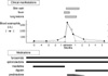
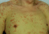
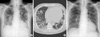
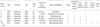
An 82-yr-old man was admitted to the hospital with a fever and cough. The patient had been diagnosed with an arrhythmia associated with dilated cardiomyopathy 9 months earlier and had been taking mexiletine for 5 months in addition to furosemide and spironolactone, which was continued since the diagnosis. He developed a fever, cough, and sputum 1 week before admission (Fig. 1). He was a retired pharmacist who lived in downtown Seoul, Korea. On physical examination, variable-sized, fused erythematous macules and plaques covered his entire body, including the trunk and extremities; this rash developed on the day of admission (Fig. 2). His body temperature was 38.3℃, and his respiratory rate was 22 times per min. Auscultation of the lungs disclosed diminished breath sounds throughout both lungs, with crackles at the bases. Laboratory studies showed leukocytosis (13,700/µL) with eosinophilia (3,310/µL; 24.2% of the white blood cells) in the peripheral blood. Liver dysfunction was also detected (aspartate transaminase 193 U/L; alanine transaminase 321 U/L, lactate dehydrogenase 290 U/L). Chest radiographs showed multiple increased opacities with a patchy distribution in both lungs (Fig. 3A). Chest computed tomography (CT) on admission showed multiple nodular consolidations with ground-glass density in both hemithoraxes and multiple mediastinal lymphadenopathy (Fig. 3B). A fine needle aspiration biopsy of a lung lesion was performed and showed eosinophilic infiltration with histiocytes, and granular pneumocytes with an organizing alveolar exudate (Fig. 4A). In addition, a right abdominal skin lesion was biopsied. The dermis showed extravasated red blood cells and moderate perivascular infiltration of lymphocytes and eosinophils (Fig. 4B). After admission, he was diagnosed with type II diabetes mellitus for the first time based on a fasting serum glucose of 183 mg/dL and hemoglobin A1c of 8.6%. The patient was diagnosed with drug-induced hypersensitivity syndrome based on the clinical findings and laboratory evidence. The mexiletine was suspected to cause theses adverse reactions and was withdrawn, while furosemide and spironolactone was continued. For the treatment of severe immune response, prednisolone 0.5 mg/kg was given for 10 days and tapered. Subsequently, his temperature decreased, with resolution of the blood eosinophilia, liver dysfunction, and cutaneous and lung lesions (Fig. 3C). With the resolution of adverse reactions, digoxin was susbstituted for mexiletine to treat arrhythmia.
Four months after resolution of the adverse findings, a patch test was performed to confirm the etiologic diagnosis. Mexiletine (Mexitil®, Boehringer Ingelheim Korea, Seoul, Korea), furosemide (Lasix®, Handok Pharmaceuticals Co., Seoul, Korea), and spironolactone (Aldactone®, Pharmacia Ltd., UK) at concentrations of 0, 1, 2, 5, 10, and 20% in petrolatum was applied at the back of the patient using Finn chambers on Scanpor tape for 48 hr. At 30 min and 48 hr after removal of the tape, the responses were scored according to the International Contact Dermatitis Research Group guidelines (9). Weakly positive reactions were detected with 5, 10, and 20% mexiletine, while ten control subjects showed negative result to the test. Based on clinical course and patch test, we were able to identify mexiletine as the etiologic agent for the development of drug-induced hypersensitivity syndrome. To explore the association of these events with human herpes virus 6 (HHV-6) infections, we performed polymerase chain reaction (PCR) analysis for HHV-6 in the serum of the patient taken at the time of patch test. The PCR analysis did not detect HHV-6 DNA.
DISCUSSION
Drug-induced hypersensitivity syndrome is the name for the severe adverse reactions to drugs as a result of systemic immune responses and is often called DRESS (drug reaction with eosinophilia and systemic symptoms) (10). Anticonvulsants such as carbamazepine and phenytoin are the most common etiologic agents causing drug-induced hypersensitivity syndrome (11). In addition, allopurinol, sulfonamides, dapsone, and minocycline can cause these reactions. Mexiletine has been reported to cause severe hypersensitivity reactions, including drug-induced hypersensitivity syndrome (or DRESS). Although mexiletine is prescribed worldwide, all previously reported cases of mexiletine-induced hypersensitivity syndrome have been Japanese, suggesting a genetic predisposition (Table 1). The present case is the first report in a non-Japanese population. Despite ethnic differences, the Korean and Japanese populations share much the same genetic background, and the exclusive incidence of mexiletine-induced hypersensitivity syndrome in Far Eastern countries implies that genetic susceptibility underlies its pathogenesis. While genetic variants associated with the development of drug induced hypersensitivity syndrome are not well defined, the human leukocyte antigen HLA-B*1502 is reported to be significantly associated with the development of carbamazepine-induced Stevens-Johnson syndrome (SJS) and toxic epidermal necorlysis (TEN) (12). This genetic association may be ethnicity specific, in that strong association is observed in Han-Chinese, not in Caucasians (13). These finding can explain relatively higher incidence of the event in Han-Chinese than in Caucasians (12, 14). Like carbamazepine-induced SJS and TEN, high incidence of mexiletine-induced hypersensitivity syndrome in Far Eastern countries maybe due to ethnicity specific genetic predisposition. In addition to ethnic characteristics, all reported cases have developed in relatively old males, which suggests a sex and age predilection.
Internal organ involvement is a specific finding of hypersensitivity syndrome, and the specific organ involvement often depends on the etiologic drug (1). Allopurinol-induced reactions frequently involve the kidney, whereas phenytoin and dapsone often cause liver dysfunction. The previous reports of mexiletine-induced hypersensitivity syndrome did not reveal the specific organ involvement, except for liver dysfunction (Table 1). Ours is the first report of lung involvement in mexiletine-induced hypersensitivity syndrome. In this report, we proved eosinophilic lung infiltration with not only imaging studies but also microscopic examination of the lung lesions. In many cases of drug-induced hypersensitivity syndrome, a clinical response develops 3 weeks to 3 months after starting a medication. This was also true for mexiletine-induced cases (Table 1), except for that reported by Yagami et al. (8) and the present case. In these latter two cases, the patients developed clinical manifestations after 5 or 6 months of medication, and patch tests proved mexiletine was the causative agent.
The mechanism for the immunogenicity of these drugs is not clear, although a role of reactive drug metabolites in initiating an immune response via hapten formation has been suggested (15). Recently, the involvement of viral infection in the pathogenesis of drug-induced hypersensitivity syndrome has been explored intensively. Although we did not confirm the viral association in this case, some previous case studies of mexiletine-induced hypersensitivity syndrome have suggested a relationship between human herpes virus 6 and the development of systemic immune responses (6-8).
In conclusion, we report the first case of mexiletine-induced hypersensitivity syndrome with lung eosinophilic infiltrates. This is also the first report of these reactions in a non-Japanese population.

 XML Download