

 PDF
PDF ePub
ePub Citation
 Print
Print


INTRODUCTION
Blunt traumatic aortic rupture (TAR) has traditionally been treated with early open surgical repair. Recently, endovascular stent-grafting of TAR has been described as a less invasive and possibly more favorable option.
CASE REPORT
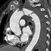
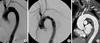
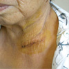
A 73-yr-old woman presented with multiple trauma and chest pain after a traffic accident. Traumatic aortic injury was suggested by a chest radiography, which showed mediastinal widening and left pleural effusion. A spiral computed tomography (CT) scan revealed an extraluminal hematoma in the distal aortic arch with a contained aortic rupture (Fig. 1). Further examination showed no other aortic lesions. However, the patient had concomitant comminuted fractures in the right tibia, left femur, and the radius bilaterally, which required surgery. Surgery for the TAR was therefore delayed for about 1 month to allow orthopedic postoperative recovery. To allow a minimally invasive surgery, a hybrid aortic stent-grafting procedure was performed, consisting of a left subclavian artery (LSCA) debranching procedure combined with proximal descending thoracic aortic stenting. Median sternotomy for the debranching procedure was avoided by transposing the LSCA to the left common carotid artery (LCCA) through a 5 cm left supraclavicular incision. The LSCA and LCCA were dissected and freed from their surrounding attachments after moderate systemic heparinization with an intravenous (IV) of 5,000 IU. The proximal LSCA was clamped and transected, and the proximal stump was closed with a continuous running suture of 4-0 polypropylene. The LCCA was partially clamped and opened longitudinally, and the transected distal portion of the LSCA was attached in an end-to-side manner. The wound was closed in layers. The patient later recovered full neurologic function without deficits. On postoperative day 5, the interventional radiologist inserted a thoracic aortic stent percutaneously using the Seldinger technique. The common femoral artery was punctured under local anesthesia. A 5F-calibrated angiographic catheter was advanced into the aortic arch via the contralateral common femoral artery to determine the morphology and extent of the aortic rupture (Fig. 2A). The delivery system was advanced under fluoroscopy after IV injection of 5,000 IU heparin until the tip reached the origin of the LCCA. Finally a SEAL® endoprosthesis (S & G Biotech, Korea) was inserted into the aortic arch to complete the procedure. A series of aortograms were performed during deployment to ensure accurate proximal and distal positioning. The proximal bare stent was placed precisely in the middle of the ascending aorta, allowing free blood flow to both arch vessels. Intraoperative angiography was used for post-procedural assessment of stent-graft positioning and endoleak (Fig. 2B). An in-hospital thoracic CT scan on postoperative day 12 (Fig. 2C) showed successful exclusion of the rupture site, with no any signs of endoleak or graft-related complications. At the first visit to the out-patient department, the patient was doing well and showed good healing of the supraclavicular incisional wound (Fig. 3).
DISCUSSION
Emergency open thoracotomy for traumatic aortic ruptures is associated with a peri-procedural mortality rate ranging between 17% and 21%, and a paraplegia rate as high as 7% (1, 2), due mainly to the large proportion of patients who present with multiple concomitant co-morbid conditions. Although delaying surgical repair of TAR may overcome the high mortality and morbidity risks associated with emergency surgery, delaying open surgery has been found to have an in-hospital mortality risk of 2-5% (3, 4). Recent results have suggested that endovascular stent-grafting may be a viable, 'minimally invasive' alternative, which can achieve the same results as emergency open thoracotomy but with lower mortality and morbidity risks (5-7). Endovascular stent-grafting, however, has several drawbacks that may limit its broader application. For example, most TARs occur at the aortic isthmus level, so the proximal landing zone in these patients is usually short. Furthermore, obligate coverage of the LSCA ostium is often required to achieve complete sealing of the rupture site, so it may cause ischemia or thrombosis (8, 9). Proximal fixation was enhanced in our patient by deploying a graft with an extended bare proximal portion. This approach could ensure adequate anchorage without aortic arch branch occlusion, even in patients with short landing zones. Furthermore, by transposing the LSCA onto the LCCA through the small supraclavicular incision, a full sternotomy could be avoided while preserving blood flow to the LSCA. Occlusion of the LSCA during unprotected stent insertion is frequently regarded as inconsequential.
In summary, this report presents our first experience with hybrid TAR, combining supra-aortic branch transposition with endovascular stent-grafting. Currently available techniques for stent-grafting of the descending aorta, combined with minimally invasive debranching procedures, may lead to the further development of safe and effective strategies that will avoid the high risks associated with conventional open surgery.
 XML Download