

 PDF
PDF ePub
ePub Citation
 Print
Print


INTRODUCTION
Penile necrosis is a rare but serious side effect of treatment with warfarin. A number of penile and skin necrosis cases have been reported in association with a familial or acquired deficiency of protein C and protein S (1-3). As initially determined in patients with hereditary thrombophilia, the proposed theoretical mechanism of warfarin-induced skin necrosis (WISN) is that necrosis occurs during a transient prothrombotic state due to the short half-life of the natural anticoagulant protein C (half-life 6 hr) compared to the half-life of the major procoagulant factor prothrombin (half-life 72 hr) (4-6). Despite this proposed mechanism, most patients with WISN have no identifiable hereditary hypercoagulable state. Given the frequent concomitant use of heparin products during warfarin initiation and the increasing recognition of heparin-induced thrombocytopenia (HIT) as an acquired hypercoagulable state, we believe that unrecognized HIT may underlie many cases of WISN.
In this report we describe a patient who developed profound necrotic penile lesions in the hypercoagulable mileu of metastatic adenocarcinoma and HIT while receiving systemic anticoagulation with low molecular weight heparin and warfarin.
CASE REPORT
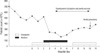
A 56-yr-old man with lung adenocarcinoma presented with subsegmental pulmonary thrombosis on April 27 2009. At the time of the first general physical examination the disease was at an inoperable stage due to multiple bone metastases. The CT scan of lungs revealed thrombosis of subsegmental and lobar pulmonary arteries within the right lung and left lower lobe. The platelet count on presentation was 531×109/L. After the diagnosis was made, the patient was started on anticoagulation with subcutaneous low molecular weight heparin (LMWH, Enoxaparin sodium, CLEXANE®) 50 mg twice daily. The next day, oral anticoagulation was initiated with 5 mg of warfarin once daily; the LMWH was discontinued on the third hospital day. On the third day of oral anticoagulation therapy, the patient complained of left leg swelling and a prolonged painful penile erection of 24 hr-duration. In addition, he was unable to void and required urethral catheterization to empty his bladder. The platelet count reached a nadir of 164×109/L at that time. There was no familial history of thrombophilia (Fig. 1). Physical examination revealed a cachectic, anxious, and severely ill man. The left lower limb was swollen, tender, and warm with tense shiny skin. There was a 13-cm difference between the circumference of the right and the left limbs above and below the knees. The penis was markedly engorged and extremely tender.
A complete blood count showed that the hemoglobin was 5.4 g, hematocrit 16.7% and the white blood cell count was 15,990 (neutrophils 87.9%, lymphocytes: 5.6%, monocytes: 5.1%, eosinophils: 1.3%, basophils: 0.1%). The prothrombin time was 61 sec (control: 12.5 sec) and international normalized ratio (INR) was 6.01. Doppler sonography of the left leg showed a deep vein thrombosis. The warfarin was discontinued and a detailed coagulation pathway analysis including protein C and S levels in the sera were performed. The protein C and S levels were assayed with functional protein C (Staclot ProteinC kit; Diagnostica Stago, Asniens-Sur-Siene, France) and radial immunodisffusion (RID), (Human Protein S 'NL' NANORID™, RID kit; The Binding Site Ltd, Birmingham, UK). The results showed that the patient had a protein C and S deficiency, with an activity level of 16% and 20% of normal values.
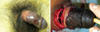
The patient was treated with narcotic analgesia in the form of 10 mg Meperidine HCl (Pethidine) intravenously, and LMWH (Enoxaparin sodium) (CLEXANE®) 50 mg twice daily. He underwent penile aspiration and blood gas analysis of cavernosal blood revealed severe acidosis, suggesting low flow priapism (pH 6.51, partial pressure of carbon dioxide 89.8 mmHg). The blood clots were extracted and irrigation with normal saline and ephedrine was performed. The next day, the left leg edema and penile erection resolved, but the penile skin became darkly discolored (Fig. 2A). On the penile Doppler ultrasonography, the penile arterial flow was intact. Seven days after the initial event, the physical findings showed necrosis of the distal half of the penis including the glans penis; a partial penectomy was performed (Fig. 2B). The pathology demonstrated hemorrhagic necrosis with organizing thrombi. Three weeks after the surgery, the patient died of complications and progression of the metastatic lung adenocarcinoma.
DISCUSSION
Bleeding is the most common complication of warfarin treatment. However, paradoxical thromboembolic events including the purple toe syndrome, skin necrosis, purpura fulminans, priapism, and penile necrosis have been reported (7). In early descriptions of WISN, patients demonstrated abnormalities of natural anticoagulant pathways, such as congenital deficiency of protein C, protein S, or antithrombin III (3-6). However, as observations of WISN accumulated, it became clear that many patients had no identifiable hereditary hypercoagulable disorder. Srinivasan et al. (8) published a series of six patients with known HIT that developed either skin necrosis or venous limb gangrene during initiation of warfarin therapy. From this case report, it is clear that the finding of skin necrosis during systemic anticoagulation with heparin overlapping with warfarin should alert one to the possibility of underlying HIT. In the current report, the penile arterial flow was intact on penile Doppler ultrasonography.
The pathogenesis of anticoagulation induced necrotic skin has been studied in patients with necrotic skin lesions due to HIT. In a series of HIT patients with venous limb gangrene, Warkentin et al. (9) reported that patients with HIT that developed venous limb gangrene were more likely to be receiving warfarin at the time of limb ischemia than other HIT patients. In addition, these patients had a significantly higher INR at the time of the development of gangrene than HIT patients that only had arterial and/or venous thrombotic events. However, despite the elevated INRs, these patients had higher levels of thrombin-antithrombin complexes simultaneously. Coagulation factor studies have revealed that these patients have severe depletion of factor VII (the coagulation factor most rapidly affected by vitamin K antagonists) as well as protein C. These observations suggest that patients with hypercoagulable disorders are predisposed to skin necrosis during anticoagulation administration due to an increase in the ratio of thrombin-antithrombin complexes to protein C activity (10, 11).
Penile ischemia and necrosis can be caused by penile prosthesis, severe diabetes, thrombotic phenomena and a buildup of calcium deposits in patients on dialysis. Acute ischemia of the penis is rarely seen because of the excellent collateral circulation of the perineum and lower abdomen (12). There are at least three mechanisms for acute ischemia of the male genitalia-widespread ligation of collateral circulation to the penis, spontaneous embolism and operative embolism (13). Other forms of venous infarction can occur from priapism and intracorporeal injection of pharmacotherapeutic agents (13). Gradual necrosis has been seen with low cardiac output and renal failure (12).
Priapism is a medical emergency. It is defined as a persistent penile erection that continues hours beyond, or is unrelated to, sexual stimulation (14). Our patient had no history of trauma or neurological disease. Priapism occurred in association with lower limb deep vein thrombosis and anticoagulation therapy. Priapism in patients with protein C deficiency alone or in association with FV Leiden has been rarely described (15, 16). Anticoagulants such as heparin, warfarin and acenocoumarol have been implicated in the etiology of priapism (16).
In the current report, the present patient developed penile necrosis with priapism that was initially attributed to WISN alone. However, the degree of penile necrosis was severe, and the patient had relative thrombocytopenia at the time of necrosis; we could not rule out HIT in this case. As this patient illustrates, clinical evidence of HIT may be evident before the development of thrombocytopenia or a 50% relative drop in the platelet count. Moreover, our patient had protein C and S deficiency. Thus, the patient's penile skin necrosis can be attributed to the underlying hypercoagulable milieu of HIT, warfarin treatment, and metastatic adenocarcinoma. As this case illustrates, it may be initially unclear whether anticoagulation-induced skin necrosis is due to WISN or a manifestation of a hypercoagulable disorder such as HIT. Therefore, HIT should always be considered in the differential diagnosis of anticoagulation-induced skin necrosis given the frequent concomitant administration of heparin products with warfarin and the increasing recognition of HIT as one of the most common acquired hypercoagulable disorders.
Treatment of HIT requires anticoagulation with one of two classes of anticoagulation agents. Direct thrombin inhibitors or heparinoids. Three direct thrombin inhibitors are currently available for patients with HIT: lepirudin, argatoban, and bivalirudin. These agents directly bind and interact with thrombin, and unlike heparin, do not require antithrombin. Direct thrombin inhibitors have short half-lives and show no cross-reactivity with heparin (17). However, we cannot use direct thrombin agents or heparnoids, because these agents have not been introduced in Korea; therefore, we waited until one day after the platelet count reached its nadir and then started LMWH. After treatment with the LMWH, the platelet counts increased and reached a stable plateau.
Given the rare, but well-documented risk of cutaneous necrosis during warfarin treatment in patients with HIT, several precautions should be undertaken to minimize the risk of skin necrosis. First, unopposed warfarin administration should be avoided in these patients and adequate levels of an alternative anticoagulant should be ensured prior to treatment with warfarin (17). The alternative anticoagulant should not be discontinued until the platelet count reaches a stable plateau and until there is at least five days of overlap with the INR>2 for two consecutive days (17). In addition, excessive levels of warfarin anticoagulation should be avoided in order to prevent a precipitous decrease in protein C levels (11). As was mentioned earlier, a supertherapeutic INR during warfarin initiation in a thrombophilic patient does not necessarily guarantee protection against thrombin generation.
Because of the early effects on protein C, warfarin can precipitate penile necrosis in the extreme hypercoagulable milieu of HIT. In patients with HIT, unopposed warfarin should be avoided. Warfarin should be initiated at modest doses in patients with HIT after platelet recovery.
 XML Download