

 PDF
PDF ePub
ePub Citation
 Print
Print


INTRODUCTION
Bisphosphonate is the treatment of choice for osteoporosis, Paget's disease, hypercalcemia of malignancy and some other skeletal complications of malignancy. Bisphosphonates are synthetic analogues of inorganic pyrophosphate (PPi) and consist of two phosphonate groups linked by nonhydrolysable phosphoether bonds to a central carbon atom, which is also attached to two covalently bonded side chains (R1 and R2). This backbone structure enables bisphosphonates to bind strongly to divalent metal ions such as Ca2+ at osteoclastic resorption sites and inhibit osteoclast function (1). Bisphosphonates are classified into two groups based on their modes of action. First generation bisphosphonates, which do not contain nitrogen, form non-hydrolyzable ATP analogues (2) and disrupt osteoclast function by being incorporated into toxic non-hydrolyzable adenosine triphosphate analogs. On the other hand, new generation bisphosphonates, which have a nitrogen containing side chain, inhibit the farnesyl diphosphonate synthase, a component of the cholesterol mevalonate pathway, and thereby disrupt cell signaling and cause apoptosis (3).
Despite their comparative efficacies, second generation bisphosphonates have well established toxicities, such as, fever accompanied by mild flu-like symptoms like fatigue, myalgia and arthralgia (4). These side effects are specific to new generation bisphosphonates and typically occur in about one-third of patients administered aminobisphosphonate for the first time (4). Furthermore, clinical features of pyrexia with transient decreases in leukocyte and lymphocyte counts and increases in CRP levels are highly suggestive of an acute phase response (4-6).
Recent progress has elucidated the mechanism underlying acute phase response induced by aminobisphosphonate. Published data suggested that the stimulations of proinflammatory cytokines, such as, IL-6 and tumor necrosis factor α (TNF-α), played a key role in pathogenesis of bisphosphonate induced fever (6-8). And Santini et al. (9) suggested that interferon γ (IFN-γ) was involved in the pathogenesis of acute phase response and in the antiangiogenic action of bisphosphonate in cancer patients.
Both alendronate and pamidronate are the aminobisphosphonates. Alendronate is either given daily or weekly for a month while pamidronate is usually administered every 3 months intravenously.
Several studies have been conducted on acute phase response to aminobisphosphonate but they were done at doses for malignancy (6-8). No study was performed regarding how the aminobisphosphonate affected patients at the dose usually used to treat osteoporosis. Therefore, the aim of the present study was to investigate the acute-phase response in patients with rheumatism after administration of aminobisphosphonate at the dose for osteoporosis.
MATERIALS AND METHODS
In vitro study: Blood sampling and isolation of peripheral blood mononuclear cells (PBMCs)
Twelve osteoporotic patients were included and the diagnosis of osteoporosis was met the ISCD guideline (10). After obtaining an informed consent, venous blood (15 mL) samples were drawn in the heparin containing syringe.
After centrifugation, the blood cells were diluted 1:1 with RPMI 1640 (Hyclone, Logan, UT, USA) wash media containing 2% fetal calf serum, layered over the Histopaque (HISTOPAQUE-1077, Sigma, St. Louis, MO, USA) and centrifuged at 693 g for 30 min at 25℃. In each case, PBMC layer at inter-phase between plasma and Histopaque was collected and washed twice in RPMI 1640. Cell pellets obtained by centrifugation were resuspended in RPMI1640/FCS. The number of PBMC in the suspensions was counted using a hemocytometer.
RNA extraction and reverse transcription
Cell pellets of 1×10⁶ cells were used for each RNA expression. Cell pellets were lysed by 1 mL of Trizol reagent (Invitrogen, Carlsbad, CA, USA) to prepare total RNA. The cDNA was synthesized using a reverse transcription system (AMV reverse transcriptase, Promega, Madison, WI, USA).
Culture of mononuclear cells
PBMC (5×10⁵) were cultured in the 96 well plate in RPMI 1640, supplemented with 2 mM glutamine and 100 IU/mL of benzyl penicillin, streptomycin (100 µg/mL) (Invitrogen, Carlsbad, CA, USA), 10% heat-inactivated fetal bovine serum (FBS) (Hyclone), and human M-CSF, Macrophage colony-stimulating factor, (Sigma) in a CO2 incubator at 37℃. Every 4-5 days 50% of the media were changed.
Incubation (pulse) of the PBMCs with alendronate
PBMC were incubated with or without alendronate (100 µM) for about 18 hr in separate culture tube and washed. PBMC were either cultured further or RNA was extracted from the cells to determine the short-term effect of bisphosphonate. 1×10⁶ cells were harvested for RNA extraction.
mRNA expressions of the inflammatory cytokines in the PBMC
The sequences of the RT-PCR primers and the real time RT-PCR probes used are shown in Table 1. The real-time RT-PCR was performed using an iCycler iQ Real-Time PCR Detection System (Bio-RAD, Hercules, CA, USA). The amplification reactions were performed in 25 µL final volume containing 12.5 µL of iQ Supermix (Bio-RAD), 1 µL of primer, 2 µL of TaqMan probe, 1 µL of cDNA, and 7.5 µL of distilled water. Thermal cycling conditions were; 10 min at 95℃ followed by 50 amplification cycles of 30 sec at 95℃ and 1 min at 60℃. Real time RT-PCR CT value were calculated using computer software (iCycler, Bio-Rad). Results were analyzed by 2-ΔΔCT relative gene expression quantification method using GAPDH as an internal standard (11). Samples were processed in duplicate.
Patients for in vivo study
Twenty-six osteoporotic patients were included in the study. Inclusion/exclusion criteria were as follows. Patients were osteoporotic (BMD<T score -2.5), but without history of bisphosphonate administration. In addition, patients were required to have at study entry, neutrophil count ≥1.5×10⁹/L, electrolytes within the normal range, normal hepatic and renal function, and no acute or chronic infections as reflected by a normal serum C-reactive protein (CRP) level (<0.3 mg/dL). Patients were considered ineligible if they had received chemotherapy or immunosuppressant during the previous 6 months, or had taken steroid dose equivalent to more than 10mg of prednisolone within previous 1 week. This study was conducted after obtaining Inha university institutional review board approval (No. 2006-201) and informed consent was obtained from all patients.
In vivo study design
This was an open, prospective study about the acute effects of pamidronate (intravenous, 30 mg in 500 mL of 0.9% saline over 3 hr) including the acute phase inflammatory response. The acute phase reactants, erythrocyte sedimentation rate (ESR) and CRP were measured just before the treatment and 24, 48 and 72 hr afterwards. Venous blood were examined for inflammatory cytokine levels (TNF-α, IL-6 and IFN γ), white blood cell (WBC) count, absolute neutrophil count (ANC), monocyte count and for bone tunrover markers including serum calcium, serum c-telopeptide (CTX). The blood for osteoclast culture was drawn into EDTA tubes before and after 48 hr after pamidronate infusion. For serum cytokine determinations, blood was rapidly centrifuged and the serum was kept at -70℃ before analysis.
Analysis of parameters
Serum levels of IL-6, TNF-α, IFN-γ and CTX were measured with enzymeimmunometric assay kits (serum IL-6 and TNF-α, R&D Systems, Minneapolis, MiN., USA; serum IFN-γ, eBioscience, San Diego, CA, USA; serum CTX, Nordic Bioscience Diagnostics, Bolden, UK). The mean minimum detectable concentrations of IL-6, TNF-α, IFN-γ and CTX were 0.039 pg/mL, 0.106 pg/mL, 4 pg/mL and 0.020 ng/mL, respectively. Samples were analyzed in duplicate. ESR was measured with the Westergren method, and serum CRP was determined by latex agglutination using a Hitachi 747 (Hitachi, Tokyo, Japan).
Cytochemical assessment of osteoclast formation
Venous blood samples were diluted 1:1 in a-MEM, layered over Histopaque-1077 and centrifuged at 711 g for 30 min. The PBMC layers were collected and washed in a-MEM. PBMC (1×10⁶ cells/well) were placed in a 96-well tissue culture plate and cultured for 14 days in a-MEM containing 10% serum, 100 units/mL penicillin, 100 µg/mL streptomycin, 50 ng/mL of M-CSF (Macrophage colony-stimulating factor) and 100 ng/mL of receptor activator for nuclear factor κB ligand (RANKL). The cells were incubated at 37℃ in a CO2 incubator. After 14 days of culture histochemical staining for tartrate resistant acid phosphatase (TRAP) was carried out using an acid phosphatase kit (Sigma 386-A) according to the manufacturer's instruction. TRAP positive giant cells with more than 3 nuclei were regarded as osteoclasts. The numbers of the osteoclasts were expressed as the counts per well.
Statistical analysis
Data are expressed as mean±SD (Standard Deviation). The mRNA expressions of the cytokines which were measured by real time RT-PCR were analyzed by Wilcoxon's test for nonparametric dependent variables.
Serum cytokine levels and the number of osteoclasts in samples cultured before treatment (basal value) were also compared with those detected in PBMCs at 48 hr after drug infusion using Wilcoxon's test for nonparametric dependent variables. Determinations of acute phase reactant levels such as ESR and CRP before and at day 1, 2 and 3 days after treatment were carried out using repeated measure ANOVA. Spearman rank correlation coefficients were used to examine the correlation between cytokine levels and CRP. Significance was accepted for P values of <0.05.
RESULTS
The demographic characteristics of patients enrolled both in vitro and in vivo studies are described in Table 2.
In vitro expression of inflammatory cytokines after alendronate incubation
After incubating PBMCs with alendronate for 18 hr, the mRNA expressions of inflammatory cytokines increased; IL-6 mRNA expression increased most prominently among them. In addition, IFN-γ also showed a significant increase. The TNF-α mRNA expression was also elevated, but the IL-1 mRNA expression did not change significantly compared to pre-incubation level (Table 3).
Expression of bone metabolism-related markers after incubation with alendronate
The mRNA expression of cathepsin K increased after incubation with alendronate, but other osteoclastogenic cytokines showed no significant change. The mRNA expression of RANKL and TRAP increased marginally and the mRNA expression of receptor activator for nuclear factor κB (RANK) showed a slight change compare to that of baseline (Table 3).
Acute phase reactants after in vivo pamidronate infusion
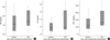
A continuous increase in serum CRP levels was observed during three consecutive days after treatment (Fig. 1). Mean basal CRP was 0.13±0.10 mg/dL and this rose to 0.26±0.30 mg/dL on day 1, to 0.55±0.73 mg/dL on day 2 and to 1.33±2.70 mg/dL on day 3, and these increases were significant (P=0.026). However, increases observed in ESR levels were not significant (P=0.312).
Inflammatory cytokine synthesis after in vivo pamidronate infusion
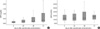
All serum inflammatory cytokine levels were significantly elevated at 48 hr after pamidronate infusion (Fig. 2). Specifically, TNF-α increased from 1.89±2.51 pg/mL at baseline to 2.33±2.78 pg/mL (P=0.009); and IL-6 increased from 1.98±1.72 pg/mL at baseline to 3.94±4.62 pg/mL (P=0.014). Increase of IL-6 levels were found to be correlated with those of CRP (P=0.040). IFN-γ increased from 20.56±32.99 µg/mL at baseline to 21.40±13.29 µg/mL at 48 hr after administration and the increase was also significant (P=0.035).
Changes in bone turnover markers after pamidronate infusion
The serum levels of bone turnover markers showed a rapid decline after pamidronate infusion (Table 4). Serum CTX which was 0.47±0.33 ng/mL at baseline, decreased to 0.14±0.10 ng/mL on day 2 (P<0.001), and serum calcium, which was 9.31±0.32 mg/dL at baseline, dropped to 8.98±0.47 mg/dL at 48 hr after the drug administration (P=0.020).
Effect of pamidronate on hematologic parameters
Hematologic parameters, namely WBC, ANC and lymphocyte counts decreased slightly. Monocyte counts were unchanged by pamidronate infusion (Table 5).
Effect on osteoclastogenesis
TRAP positive cells with more than three nuclei were regarded as osteoclasts. The number of TRAP positive giant cells formed after culturing PBMCs were 47.1±43.9 cells/well at baseline, and this decreased to 37.6±51.1 cells/well on day 2 but this reduction was not statistically significant (P=0.080).
DISCUSSION
Our preliminary in vitro study on the effect of alendronate (100 µM) on PBMCs after incubation for 18 hr showed that the mRNA expressions of the inflammatory cytokines such as IL-6, TNF-α and IFN-γ increased. This finding indicates that mRNA expressions of the inflammatory cytokines occur as early as 18 hr after the administration at the cell level while the symptoms of acute phase response occur 24-36 hr after administration (4).
The subsequent in vivo study was undertaken to determine whether pamidronate infusion affects the productions of these inflammatory cytokines after administrating a usual dose of pamidronate at 30 mg. It was found that pamidronate significantly increased both serums IL-6 and TNF-α levels in vivo, which concurs with the findings of previous studies (7, 8).
As acute phase reactant, CRP levels were found to increase steadily over three consecutive days following pamidronate infusion. IL-6 has been known to play a key role in the stimulation of synthesis of CRP by the hepatocytes (12) and it was interesting to find in the present study that increment in IL-6 and CRP significantly correlated each other.
The mechanism for acute phase response and the identities of the effector cells that release IL-6, TNF-α and IFN-γ have been recently reported (13). During the inhibition of the mevalonate pathway by aminobisphosphonate, the metabolic intermediates, such as, isopentenyl pyrophosphate (IPP) accumulated. IPP was a potent activator of human peripheral blood γδ T cells (14). When activated, the γδ T cells rapidly produced large quantities of IL-6, TNF-α and IFN-γ (13, 15). During a 'typical' acute phase response, monocytes and macrophages were major source of the cytokines but during acute phase response to bisphosphonate, human γδ T cells produced these proinflammatory cytokines (13), activated by bisphosphonate as the antigen presented on the surface of monocyte lineage cells (15). The potencies of aminobisphosphonates appeared to correlate inflammatory milieu. The aminobisphosphonates with stronger potencies increase more IPP production, which led to the activation of larger numbers of peripheral blood γδ T cells and as such, more proinflammatory cytokines were induced (16).
The intermittent large dose bisphosphonate for cancer treatment increased bioavailability and also the incidence of acute phase response (17). This seemed to be the reason why many studies on acute phase response were carried out at the dose for treatment of cancer (7, 8, 18-20). In these studies, transient pyrexia was found to be associated with increases in inflammatory cytokines, whereas in our patients elevated temperatures were rarely observed (data not shown). One Korean study reported that acute phase response occurred in 55.6% of osteoporotic patients after administration of intravenous pamidronate but the symptoms were mostly mild such as myalgia and febrile sense persisting for less than 2 days (21). Therefore we tried to figure out whether bisphosphonate affected inflammatory cytokines in osteoporotic patients. And it was confirmed in this study that inflammatory cytokines and acute phase reactants were significantly increased regardless of elevation of body temperature. The dose of pamidronate required to treat osteoporosis was sufficient to cause an acute phase response within 48 hr after the administration although it barely affected hematopoietic parameters. Sauty et al. (7) found significant decreases in lymphocyte and leukocyte counts after a 60 mg pamidronate infusion 48 hr afterwards, which was twice the dose used in our study.
Nevertheless, even at the dose which caused barely no change on hematologic parameters, pamidronate was found to affect bone metabolism within 48 hr after administration. The declines in serum CTX and serum calcium at 48 hr after administration suggested that the intermittent large dose pamidronate have a rapid onset of action.
One interesting observation in the present study was that mRNA expression of IFN-γ was increased in in vitro and in vivo studies. A previous study on pamidronate 60 mg also showed elevated serum IFN-γ level in in vitro study (22) and another study confirmed this at 90 mg dose of pamidronate in vivo (9). IFN-γ is a pleiotropic cytokine that is secreted by activated CD4 and CD8 T cells and has immunomodulatory effect. Some evidences indicated it had an antiangiogenic effect by inhibiting endothelial proliferation (23). One of the worst side effects of bisphosphonate, osteonecrosis of the jaw (ONJ) was known to be associated with its antiangiogenic effect (24). Despite the incidents of ONJ at the dose used to treat malignancies, bisphosphonate was regarded safe for the treatment of osteoporosis (25). However, as intermittent large dose bisphosphonate therapy is favored nowadays, the probability of ONJ would be increasing; increase in IFN-γ in our in vitro and in vivo studies supported this hypothesis.
The limitation of this study is that different bisphosphonate was used in vitro and in vivo studies. Both alendronate and pamidronate are aminobisphosphate and have very similar property. Intravenous regimen of alendronate has not been available to date and this study was performed before the introduction of intravenous ibandronate. In order to test on the hypothesis of this study, we chose the only aminobisphosphate, pamidronate, which was allowed in human use for intravenous injection. We assumed that both of them would cause similar inflammatory reactions.
In conclusion, acute phase response is noted after intravenous administration of aminobisphosphonate at osteoporotic dose. Our studies confirmed that intermittent large dose aminobisphosphonate therapy causes acute inflammation despite rapid efficacy on osteoporosis; hence we suggest that intermittent large dose bisphosphonate should be used with caution.


 XML Download