

 PDF
PDF ePub
ePub Citation
 Print
Print


INTRODUCTION
Systemic chemotherapy can lead to a variety of ocular complications, such as cicatricial ectropion, nasolacrimal duct stenosis, conjunctivitis, keratitis, cataract, macular edema, retinopathy, and optic neuropathy (1, 2). Although bulbar perforation with orbital cellulitis has been reported in an immunocompromised patient, corneal perforation has not been documented in patients undergoing systemic chemotherapy (3).
We report a case of corneal perforation with preseptal cellulitis in a patient treated with systemic chemotherapy for acute lymphocytic leukemia (ALL).
CASE REPORT
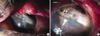
A 17-yr-old female patient undergoing systemic chemotherapy for ALL was referred to our hospital due to swelling and pain of the right upper lid for two days. Laboratory examination showed leukocytes 40 cells/µL, erythrocytes 3.6×10³ cells/µL, hemoglobin 10.8 g/dL, hematocrit 30.6%, and thrombocytes 33×103 cells/µL. The patients received induction chemotherapy (vincristine, prednisolone, daunorubicin, and L-asparaginase). Visual acuity in the right eye was 20/20. There were no abnormal findings, other than diffuse swelling of the right upper eyelid. Orbital computed tomography showed findings of preseptal cellulitis in the right upper lid. Intravenous empirical antibiotics (teicoplanin, meropenem, and amphotericin B) were administered. Serratia marcescens was isolated from blood culture on the following day. Slit lamp examination on the 7th day revealed superficial punctate erosions in both corneas. Artificial tear eye drops were used for the treatment of superficial punctate erosions. On the 12th day, spontaneous bloody and purulent discharge from the upper palpebral conjunctiva occurred in the right eye. The patient was treated with topical levofloxacin eyedrops. Culture from the discharge was isolated from S. marcescens. Systemic antibiotics (meropenem) were maintained per the antibiotic sensitivity test. On the 16th day, periorbital swelling decreased, however, corneal melting and perforation with iris prolapse was noted in the right eye (Fig. 1). Emergent tectonic keratoplasty was performed. Seven months after surgery, visual acuity in the right eye was 20/300, and intraocular pressure was 14 mmHg. The corneal graft was stable, with mild haziness.
DISCUSSION
Ocular complications associated with systemic chemotherapy can be divided into complications in the adnexa, anterior segment, and posterior segment. Previously reported complications in the anterior segment include conjunctival injection, conjunctivitis, corneal edema, keratitis, and corneal opacity (1, 2). Anticancer agents such as cytosine arabinoside, 5-flurouracil, carmustine, deoxycoformycin, and tamoxifen have been known to cause corneal toxicity (1, 2, 4, 5). In our case, vincristine, prednisolone, daunorubicin, and L-asparaginase were used for combination chemotherapy. To the best of our knowledge, corneal toxicity associated with these anticancer agents has not been reported.
Serratia marcescens, a motile, Gram-negative coccobacillus, is an emerging opportunistic pathogen noted for causing urinary, respiratory, blood stream, and ocular infections (6). Preseptal cellulitis in the present case was caused by S. marcescens because the patient was immunocompromised. Rapid progression of stromal infiltration and ulceration is a clinical feature of Serratia keratitis. Extracellular protease produced by S. marcescens is considered a major corneal destructive factor. In an experimental study of rabbit cornea, Serratia protease preparations have caused rapid and extensive liquefactive corneal necrosis, descemetocele formation, and corneal perforation (7).
The exact mechanism of corneal melting and perforation in this case is unclear. It is possible that S. marcescens in the purulent discharge directly invaded the cornea with superficial punctate erosions and caused corneal melting and perforation in the immunocompromised patient. This hypothesis is supported by two facts: First, S. marcescens is a virulent organism. Second, corneal melting and perforation occurred in the area in direct contact with the infected upper palpebral conjunctiva.
In conclusion, physicians should consider the possibility of serious ocular complications, such as severe bacterial keratitis or corneal perforation in cases of preseptal cellulitis caused by a virulent organism, particularly in immunocompromised patients.
 XML Download