

 PDF
PDF ePub
ePub Citation
 Print
Print


INTRODUCTION
In 1958, Cantrell et al. (1) described a syndrome which consists of the following: 1) a midline, supraumbilical abdominal wall defect; 2) a defect of the lower sternum; 3) a deficiency of the anterior diaphragm; 4) a defect in the diaphragmatic pericardium; 5) congenital intracardiac defects.
Several authors have described similar cases, and it is currently classified as an anterior body wall midline developmental anomaly (2). The estimated prevalence varies from one in 65,000 to 200,000 births (2, 3). In this case report, we describe successful vaginal delivery of a pregnant woman who had been diagnosed with this syndrome in childhood.
CASE REPORT
A 23-yr-old nulliparous, primigravid woman was referred to our maternal-fetal medicine unit at 18 weeks' gestation. She had been previously seen at our institution in the pediatric clinic as a child of 6 yr, when she presented with two umbilici and an epigastric pulsatile mass present since birth. At that time, she complained of intermittent abdominal pain in the area between the two umbilici. On physical examination, the superior umbilicus was found to be a dimple in the epigastrium connected to the actual umbilicus inferiorly by a fibrous band. A 1×3 cm pulsatile mass was palpable above the dimple.
Chest plain radiograph showed situs solitus and slightly decreased pulmonary vascularity and abdominal plain radiograph was unremarkable. On EKG, left ventricular hypertrophy was suspected. Cardiac catheterization, angiography, and echocardiography were performed and the pulsatile mass was found to be a substernal apical diverticulum of the heart beneath the weakened abdominal wall. Diagnosis of Cantrell's Pentalogy was made. Periodic follow-up studies over the next 6 yr, including chest radiography, EKG, and echocardiography showed no remarkable changes.
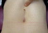
At age 23 yr, she presented to our institution at 18 weeks' gestation and was examined by echocardiography. The echocardiogram revealed left ventricular ejection fraction of 63%, suspected mild right ventricular outflow tract compression without a pressure gradient, and insignificant mild narrowing of left pulmonary artery origin. During prenatal evaluation, the patient did not complain of any significant symptoms except for a pulsatile sensation in the epigastrium and mild intermittent dyspnea. There were no abnormal obstetric findings, including antenatal routine laboratory results, midtrimester maternal serum screening test, and vaginal examination. On fetal ultrasonogram, fetal growth appeared normal, and fetal anomaly was not found. An epigastric dimple and the umbilicus were connected by a fibrous band, and the substernal cardiac diverticular beating beneath the abdominal wall was similar as previously described (Fig. 1).
At 39 weeks and 6 days' gestation, she was admitted for oligohydramnios. Induction of labor with vaginal dinoprostone insertion and intravenous oxytocin was performed. During the intrapartum period, she was monitored closely for any abnormal symptoms or signs. She delivered a 3.5 kg male baby without complication. The 1-min and 5-min Apgar Score was 9 and 9 and there was no gross anomaly noted including the umbilical cord and umbilicus. Postpartum uterine contraction was adequate, and she was discharged with the healthy baby without any significant hemorrhage or abdominal pain.
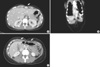
One year after the delivery, she underwent abdomino-pelvic CT. Appreciable on the CT were the apical diverticulum and pericardial defect (Fig. 2A), lower sternum and diaphragm and pericardium defect (Fig. 2B), and abdominal wall defect (Fig. 2C). She had experienced no significant problems since delivery for one year.
DISCUSSION
Previous case reports have described various conditions associated with Cantrell's Pentalogy, but as far as we are aware, this is the first reported case of successful vaginal delivery of a pregnant woman diagnosed with this condition.
All five anomalies must be present for diagnosis of Cantrell's Pentalogy to be made, but the severity may be quite variable, ranging from subtle to lethal. Prognosis depends on the severity of the cardiac and associated anomalies. In affected patients with complex heart anomalies who undergo surgery during the first days of life, mortality can be as high as 50%. Possible causes of death include cardiac rupture, tamponade, endocarditis, embolism, arrhythmia, and heart failure (3, 4).
The etiology of Cantrell's Pentalogy is unknown, but it has been postulated that a defect in the common mesodermal origin accounts for the association of the five anomalies. Most of the reported cases are sporadic, but X-linked inheritance has been suggested in some families, and mutations in genes located on the X chromosome have been suspected (5, 6). It is possible that some intestinal loops pass the umbilical ring and herniate into the cord during embryonic development, and a defect in normal internalization of the physiological umbilical hernia results in a persistent herniation and abundant umbilical skin (7).
Delivery mode has not been discussed for maternal Cantrell's Pentalogy. In fact, the care of pregnant woman with Cantrell's Pentalogy has not been previously described. With close antenatal follow-up and intrapartum monitoring, we were able to perform successful vaginal delivery. We observed the patient closely for any symptoms and signs of dyspnea, abnormal abdominal pain, or tachycardia. This case report demonstrates that vaginal delivery may be an option for women with Cantrell's Pentalogy.
 XML Download