

 PDF
PDF Citation
 Print
Print


INTRODUCTION
Nail-patella syndrome (NPS; OMIM #161200) is an autosomal dominant disorder characterized by classic clinical tetrad of dysplastic finger nails, absent or hypoplastic patellae, elbow dysplasia, and exostoses ("horns") of the ilia (1). In addition, some patients manifest nephropathy and adult-onset glaucoma (2, 3). The incidence of NPS is estimated at 1 in 50,000 live births (4). NPS is highly penetrant, but the phenotypes show marked inter- and intrafamilial variability (4).
NPS is caused by loss-of-function mutations in the LMX1B gene, which encodes a LIM-homeodomain transcription factor (5, 6). Animal studies have shown that the LMX1B gene plays an important role in dorsal-ventral patterning of limb development, morphogenesis and function of glomerular basement membrane (GBM) and podocyte, as well as in development of anterior segment of the eye (7-9). To date, more than 140 mutations of the LMX1B gene associated with NPS have been identified (The Human Gene Mutation Database at the Institute of Medical Genetics in Cardiff http://www.hgmd.cf.ac.uk/ac/index.php).
This study was conducted to evaluate the clinical manifestations of 9 unrelated Korean children with NPS and 6 of their affected parents, to investigate mutations of the LMX1B gene, and to analyze the phenotype-genotype correlation.
MATERIALS AND METHODS
Nine unrelated Korean children, who were clinically diagnosed with NPS by the Department of Pediatrics or the Department of Pediatric Orthopedic Surgery at Seoul National University Children's Hospital were enrolled in this study. A pediatric orthopedic surgeon evaluated skeletal abnormalities of the patients by physical examination and radiologic studies. The presence of glaucoma in the adult family members of the patients was evaluated based only on a review of the patient history.
Mutational analysis of the LMX1B gene was conducted for all 9 patients, and their available family members. Genomic DNA was prepared from peripheral blood nucleated cells. The 8 coding exons of the LMX1B gene were then amplified by polymerase chain reaction (PCR) and directly sequenced. The sequences of the PCR primers are shown in Table 1.
A recent study revealed that the coding sequence of the human LMX1B gene is longer than previously reported, and that it includes an additional 23 amino acids at the N-terminus; therefore, the numbering of the LMX1B mutations has been adjusted (10). However, in our study, the older numbering system was used to make the comparison with previously reported mutations easier.
This study was approved by the Ethics Committee of Seoul National University Hospital, Seoul, Korea, and informed consent for the genetic analysis was obtained from all patients and/or their parents.
RESULTS
The probands included 5 boy and 4 girls with a median age at the time of clinical diagnosis with NPS of 6.8 yr (range 1.2-13.6 yr). The presenting manifestations included symptoms associated with knee abnormalities (habitual patellar dislocation, pain, locking or clicking) in 6 patients, elbow contracture in 1 patient, nail hypoplasia in 1 patient, and generalized edema/nephrotic syndrome in 1 patient.
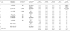
The phenotypes and genotypes of the patients and their affected family members are summarized in Table 2. Analysis of the LMX1B gene revealed 8 different mutations, including 5 missense mutations, 1 frame-shifting deletion (c.680delA) and 2 abnormal splicing mutations (IVS1-1 G>C and IVS1+5A>G). Two of the missense mutations were located in the LIM-B domain of LMX1B (p.His114Gln and p.Leu127Pro), and 3 were located in the homeodomain (p.Arg200Gln, p.Arg 200Trp and p.Ala213Pro). The p.Arg200Gln mutation was found in 2 unrelated patients. Three novel missense mutations (p.H114Q, p.L127P and p.R200W) that were identified in the probands were not detected in 100 control Korean subjects (200 alleles). In 6 of the families, one of the parents was affected both clinically and genetically; however, no siblings in any of the families were affected. Two of the patients (families 5 and 9 in Table 2) were confirmed to have a de novo mutation. Finally, the patient in family 4 was an adopted son; therefore, his family history was unavailable.
The clinical features were analyzed in 9 index cases and 6 affected parents. All patients and affected parents were found to have dysplastic thumb nails with/without the involvement of other finger nails. In addition, dysplastic toe nails were detected in 10 patients. Patellar anomalies were noted in all index cases and 4 of the affected parents. These anomalies included aplasia in 2 patients and hypoplasia in 11. Elbow contractures (cubitus valgus) with/without radial head dislocation were detected in 4 patients and 2 parents, and iliac horns were detected in 5 patients and 3 parents.
Renal involvement was detected in 2 patients and their affected parents (families 1 and 2 in Table 2). In the case of the patient in family 1, the renal involvement presented as full-blown nephrotic syndrome at the age of 2.2 yr, which did not respond to conventional oral steroid treatment and progressed to end-stage renal disease within 2 yr. A renal biopsy performed when she was 2.5 yr old revealed focal segmental glomerulosclerosis (segmental sclerosis and global sclerosis in 56% and 28% of the glomeruli, respectively). However, electron microscopic examination was unavailable due to lack of glomeruli in the specimen. Her mother had suffered from asymptomatic proteinuria since the age of 18, and had progressed to end-stage renal disease at age 28. She had also developed vestibular Schwannoma at age 26. The patient in family 2 was diagnosed with NPS at age 6, at which time mild proteinuria and microscopic hematuria were detected. Renal disease in this patient was stable until the follow-up, which occurred at age 9. Her father, who had same mutation and clinical features, developed end-stage renal disease and underwent renal transplantation at age 35.
Glaucoma or hearing difficulty was not detected in any patients or affected family members.
The range and severity of the clinical manifestations differed between and within families. For an extreme example, while the patient in family 7 had the phenotype for the entire clinical tetrad, the only phenotype shown by his mother was the absence of skin creases overlying the distal interphalangeal (DIP) joints.
DISCUSSION
NPS is caused by heterozygous loss-of-function mutations of the LMX1B gene, which encodes LMX1B, a member of the LIM-homeodomain family of transcription factors (11) LMX1B contains two cystine-rich zinc-binding motifs (LIM-A and LIM-B domain), a homeodomain, and a carboxy terminal glutamine-rich and serine-rich domain. LIM domains are involved in interactions with other transcription factors and synergistic activation of transcription. The homeodomain is involved in DNA binding, and the carboxy terminal domain represents a transcriptional activation domain (12, 13). To date, 142 LMX1B mutations have been identified and deposited in the Human Gene Mutation Database at the Institute of Medical Genetics in Cardiff (http://www.hgmd.cf.ac.uk/ac/index.php). These include 80 missense/nonsense mutations and 16 abnormal splicing mutations. Among these 80 missense/nonsense mutations, 31 (38.8%) are located in the LIM-A domain, 15 (18.8%) in the LIM-B domain, and 30 (37.5%) in the homeodomain. In our study, no mutations were detected in the LIM-A domain.
LMX1B is required for a wide range of developmental processes including dorso-ventral patterning of the limb, differentiation of dopaminergic and serotonergic neurons, patterning of the skull, and normal development of the kidney and eye (1, 5, 7, 9, 14). Accordingly, NPS shows variable phenotype with multi-organ involvement. Besides the classic clinical tetrad (dysplasia of the patellae, nails and elbows and the presence of iliac horns), other components of the musculoskeletal system such as muscle, tendons, and ligaments can be affected. In addition, other organs such as kidneys, eyes and possibly ears, the nervous system and the gastrointestinal tract can be affected as part of the syndrome (1, 4). Although NPS is a highly penetrant hereditary disorder, it shows marked inter- and intrafamilial phenotypic variability (4). In this study, autosomal dominant inheritance was confirmed both phenotypically and genetically in 7 of the 9 families, while the remaining two patients had de novo mutations. In addition, although we did not identify a genotype-phenotype correlation, we did observe inter- and intrafamilial phenotypic variability.
Changes in the nails, which were detected in all of the patients in this study, are the most constant clinical features of NPS and are detected in almost all patients. These features may include the development of only the triangular lunulae, which is one of the pathognomonic signs of NPS (15). Another common and sensitive sign of digital involvement is loss of DIP skin creases (4), which was the only abnormality detected in mother of Patient 7 in this study. Knee involvement including typical patellar hypoplasia or aplasia is also very common, and 6 of 9 patients in this study visited the hospital due to symptoms associated with their knee joints. Elbow abnormalities including limitation of joint motion, hypoplasia of the radial head, and subluxation or dislocation of the radial heads can also occur with/without antecubital pterygia. Iliac horns are bilateral and conical bony processes that project postero-laterally from the central part of the iliac bones, are considered pathognomonic of NPS. A previously conducted review of NPS reported the following frequencies of symptoms associated with the disease: nail anomalies 95.1%, patellar involvement 92.7%, elbow dysplasia 92.5%, and iliac horns 70-80% (15). Additionally, a British study of 123 NPS patients from 43 families reported that nail changes were detected in 98% of the patients, knee symptoms in 74% of the patients, elbow symptoms in 33% of the patients, and iliac horns in 68% of the patients (4). The frequencies of the abnormalities observed in our study were similar to those observed in the British study.
The incidence of renal involvement in patients with NPS has been reported to be 12-62% (4, 16), which is comparable to the results observed in the present study (27%). The earliest sign of renal involvement in NPS is proteinuria with or without hematuria, which may remit spontaneously or progress to overt nephritis. Approximately 5-15% of all patients develop chronic renal failure (4, 16). Thus, renal involvement is one of the major prognostic factors of NPS. In spite of this, the factors responsible for the development and progression of nephropathy in patients with NPS are largely unknown. However, a recent study drew the following conclusions regarding renal involvement in patients with NPS: 1) quantitative urinalysis revealed the presence of proteinuria in 21.3% of the patients, and microalbuminuria was detected in 21.7% of the patients without overt proteinuria, 2) proteinuria and microalbuminuria were found to occur significantly more frequently in females, 3) patients with an LMX1B mutation located in the homeodomain were found to have a significantly greater occurrence and higher values of proteinuria than those carrying mutations in the LIM domains, and 4) a positive family history of nephropathy and presence of radial head hypoplasia were found to be associated with an increased individual risk of developing renal disease (17). In our study, renal involvement was detected in 2 patients and their affected parents, 3 of whom were females. However, mutation located in the homeodomain and radial head hypoplasia were detected in only one of the families.
The most common renal pathologic findings characteristic of NPS are focal or diffuse and irregular thickening of the GBM with patchy electron-lucent areas (so called 'moth-eaten' appearance) and irregular deposition of bundles of fibrillar collagen (type III collagen) within the GBM and the mesangial matrix. These characteristic ultrastructural features of the GBM have been observed in all biopsied NPS patients. However, the severity of these changes do not correlate well with patient's age, the severity of proteinuria, the degree of impaired renal function, or even the presence of nephropathy (15, 16, 18-20). In our study, renal biopsy was performed in only one patient (family 1) who had developed full-blown nephrotic syndrome at the age of 2.2 yr and end-stage renal disease at age 4. Her clinical course was rather unusual because the progression to end-stage renal disease in patients with NPS is usually slow (15, 16). Her renal biopsy, which was performed at age 2.5, revealed focal segmental glomerulosclerosis. However, the GBM changes were not evaluated due to lack of glomeruli in the specimen.
Open angle glaucoma or ocular hypertension is a recently recognized phenotype of NPS (4, 6, 21). These lesions, which usually develop during adulthood, are treatable; therefore, regular ophthalmologic screening of patients with NPS should be strongly encouraged. In our study, no patients or adult family members with glaucoma were detected, but its presence was evaluated solely based on patient history.
In conclusion, the phenotypic and genotypic features of the patients evaluated in this study were similar to those observed in previously conducted studies. In addition, inter- and intrafamilial variability of the phenotypes was observed, but no genotype-phenotype correlation was observed. However, the mechanism underlying the phenotypic variations and predisposing factors of the development and progression of nephropathy in NPS patients are still unknown.

 XML Download