

 PDF
PDF Citation
 Print
Print


INTRODUCTION
IgA nephropathy (IgAN) is the most frequent primary glomerulonephritis that presents with hematuria and often proteinuria. Although a moderate degree of proteinuria is common in patients with IgAN, nephrotic syndrome is considered uncommon in these patients. Previous studies have demonstrated that some cases of IgAN presenting with the nephrotic syndrome responded to corticosteroid therapy. However, the response to steroid treatment has been variable, and usually correlated with the underlying histological changes. Some have suggested that these IgAN patients represent a variant type of IgAN (1-3); however, others have proposed that these cases might be variant type of minimal change disease (MCD), such as MCD with coincidental mesangial IgA deposits (4-6). Recently, it has been postulated that mesangial IgA deposition in MCD might represent the coincidental occurrence of MCD and IgAN (7).
We have treated several IgAN patients presenting with nephrotic syndrome. Some of these patients responded well to corticosteroid therapy, and they showed complete remission of the nephrotic syndrome. The others, however, did not respond to steroid therapy, and they had progressive renal deterioration. Since MCD is the most common cause of adult nephrotic syndrome in Korea, we thought that the steroid-responders might have concurrent MCD in addition to IgAN. In the present study, we evaluated the clinical and pathological features of patients with IgAN that presented with steroid-responsive nephrotic syndrome.
MATERIALS AND METHODS
Patients
From 2001 to 2007, a total of 581 patients were diagnosed with primary IgAN at the Seoul National University Hospital, which accounted for 37.0% of all kidney biopsies. The diagnosis of IgAN was based on immunofluorescence microscopy showing mesangial IgA deposition as the predominant or co-dominant immunoglobulin. Henoch-Schölein purpura, systemic lupus erythematosus and liver cirrhosis were excluded by detailed clinical history, physical examination and laboratory findings. Among 581 patients with IgAN, 48 patients (8.3%) presented with the nephrotic syndrome; the nephrotic syndrome was defined by generalized edema, heavy proteinuria of more than 3.5 g/day, hypoalbuminemia of less than 3.5 g/dL, and/or hypercholesterolemia of more than 200 mg/dL. Twenty five patients received steroid treatment, and a retrospective analysis revealed 12 patients that had complete remission with high-dose steroid therapy. The following blood and urinary laboratory tests were performed in all patients at varying intervals: serum creatinine, blood urea nitrogen, urine protein, and urinalysis.
A reduction of proteinuria to less than 1.0 g/day was designated as a response, and the complete correction of the biochemical findings including absence of proteinuria (less than 0.3 g/day) was considered a complete remission. Relapse of the nephrotic syndrome was defined as the recurrence of significant proteinuria (more than 1.0 g/day) and edema.
Histology grading
The light microscopy features of the renal biopsy were examined to evaluate the glomerular changes, which were classified into grades I-V according to the disease severity as described previously (8): grade I, normal or focal mesangial cell proliferation; grade II, diffuse mesangial cell proliferation, or <25% of glomeruli with crescent (Cr)/segmental sclerosis (SS)/global sclerosis (GS); grade III, 25-49% of glomeruli with Cr/SS/GS; and grade IV, 50-75% of glomeruli with Cr/SS/GS; grade V, >75% of glomeruli with Cr/SS/GS. The immunofluorescence staining was graded: 0, 1+, 2+, and 3+.
RESULTS
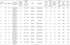
The clinical characteristics at the time of the renal biopsies are shown in Table 1. The 12 patients consisted of three males and nine females, and the mean age at the time of diagnosis was 48±18 yr. All patients complained of generalized edema, which developed acutely over two weeks (range, 1-8). The mean weight gain was 4.0 kg (% of body weight, 7.1%). Hypertension was present in four patients (33%); two patients had been diagnosed with hypertension before the renal biopsy, and the others were diagnosed at the time of the renal biopsy. Hematuria (either microscopic or macroscopic) was detected in 11 patients (91.7%), two of whom had episodes of "synpharyngitic" gross hematuria before the renal biopsy. The mean urinary protein excretion was 10.2±4.4 g/day. Two patients (16.7%) had renal insufficiency (serum creatinine greater than 1.6 mg/dL) at presentation. The mean (±SD) levels of serum albumin, serum cholesterol and serum IgA were 2.2±0.6 g/dL, 394±104 mg/dL and 285±66 mg/dL, respectively. The serum IgA level was elevated (greater than 400 mg/dL) in only one patient.
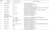
All patients had a renal biopsy at the nephrotic stage. One patient (patient 1) had already been diagnosed with IgAN on renal biopsy five years previously; due to the development of generalized edema and heavy proteinuria recently, a second renal biopsy was performed. The second biopsy showed "diffuse foot process effacement" in addition to the typical features of IgAN. The other 11 patients did not have a prior renal biopsy. The light microscopic, immunofluorescence and electron microscopic (EM) findings of the renal biopsy are shown in Table 2. The distribution of the glomerular grades of the 12 patients was: grade I, three (25%); grade II, five (42%); and grade III, four (33%). Grade IV and V IgAN was not reported among the 12 patients. There were various degrees of mesangial proliferation and abnormality. In all cases, the immunofluorescence demonstrated diffuse, granular deposition of IgA in the glomerular mesangium. Less intense fluorescence for IgG, IgM, C3, and fibrinogen were sometimes detected. There were electron dense deposits within the matrix of the mesangium and paramesangium. Subendothelial deposits (patient 8) and mesangial interposition (patient 7), were occasionally demonstrated. Extensive effacement and fusion of the visceral epithelial foot processes were detected in 8 patients (patient 4, 5, 6, 8, 9, 10, 11,and 12), while focal effacement of the foot processes were demonstrated in the remaining four patients. The glomerular basement membrane exhibited localized irregularities: thinning, lamellation and duplication (patient 3, 4, and 10).
The patients received oral prednisolone (0.5-1.0 mg/kg/day) after the renal biopsy. High-dose steroid was maintained for 4 weeks, and then it was slowly tapered. All 12 patients showed complete remission with steroid therapy. The response to the steroids was prompt; the median time to response (proteinuria of less than 1.0 g/day) was two months (range, 0-6), and the median time to complete remission (proteinuria of less than 0.3 g/day) was two months (range, 0-41). During the tapering of the steroids, six patients received other immunosuppressant; cyclosporine (3 patients), cyclophosphamide (2 patients) and azathioprine (1 patient). Patients generally tolerated the medications well with manageable side effects, including transient hyperglycemia, insomnia and cushingoid changes. There were no significant adverse events except one case of acute pyelonephritis.
The median duration of follow-up was 30 (6-84) months. Seven episodes of relapse were reported in five patients during steroid tapering. Six episodes of relapse responded to the increase of dosage or re-administration of steroids, which resulted in complete remission again. For responders, the median time to remission was two weeks (range, 0-4). Only one patient did not respond to steroid therapy; in this case mycophenolate mofetil and cyclosporine were added to the steroid treatment. At the last follow-up, the urine protein and serum creatinine were 2.27 g/day and 1.1 mg/dL, respectively, in this patient.
DISCUSSION
In this study, we evaluated IgAN associated with steroid-responsive nephritic syndrome from both the clinical and histopathological findings. It is possible that these patients had IgAN and MCD simultaneously. The clinical and pathological features of typical mesangial IgA deposits, mesangial hypercellularity and the presence of hematuria were consistent with IgAN. However, the features of wide foot process effacement on EM, sudden onset of generalized edema with weight gain, massive proteinuria, severe hypoalbuminemia, and severe hypercholesterolemia were characteristics of MCD. Rapid response to steroid therapy, steroid dependence and relapse during steroid tapering in some cases also suggested MCD.
These findings are different from those reported by Lai et al. (3), where eight patients with IgAN, presented with steroid-responsive nephrotic syndrome. They suggested that the eight patients might have a variant type of IgAN, on the basis of the clinical and pathological features including: the response to steroid treatment was not as prompt as generally observed with MCD; a higher incidence of hypertension, hematuria and azotemia was reported as compared with typical MCD cases. However, our 12 cases showed rapid response to steroid therapy; the median time to complete remission was only two months. The incidence of hypertension, hematuria and azotemia in our study was higher than that generally seen with MCD, yet the azotemia returned to normal after steroid therapy in all except one patient. Also, the mean age (48±18 yr) at diagnosis in our cases was older than that of previous studies (1, 4, 9). The old age at presentation could explain the higher incidence of azotemia and hypertension.
Interestingly, one patient already had a renal biopsy before the development of the nephrotic syndrome. The first renal biopsy showed typical mesangial IgA staining with mild mesangial proliferation, and the second biopsy, at the nephrotic stage, showed wide foot process effacement in addition to the features of IgAN. This case may represent IgAN complicated by MCD rather than a variant clinical manifestation of IgAN. The concept of these two diseases occurring simultaneously might be helpful for the management of IgAN patients with the nephrotic syndrome. The patients evaluated in this case series likely illustrated patients with the nephrotic syndrome caused by MCD in patients with preexisting IgAN, diagnosed by renal biopsy previously or not. In several cases of recurrent nephrotic syndrome (10), sequential renal biopsies have been performed with the initial biopsy showing features of MCD, not of IgAN. However, the renal biopsies obtained later, revealed features of IgAN: mesangial IgA staining on immunofluorescence, mesangial electron dense deposits on EM and/or mesangial hypercellularity. These cases showed the sequential development of MCD and IgAN during the course of steroid dependent/resistant MCD.
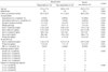
During the study period, 13 IgAN patients with the nephrotic syndrome did not respond to steroid treatment despite the same treatment protocol, and 23 IgAN patients did not received steroid treatment. We compared these 13 non-responders and 21 non-users with responders (Two patients of the non-users were excluded because of inadequate biopsy specimen.) The clinical and pathological features of these three groups are summarized in Table 3. At presentation, the responders showed greater weight gain (responders vs. non-responders; 4.0 vs. -0.3 kg; p=0.003 by post-hoc Mann-Whitney U test with Bonferroni correction), more proteinuria (10.2 vs. 4.7 g/day; p=0.003) and lower serum albumin (2.15 vs. 3.15 g/dL; p<0.001) than non-reponders. The initial serum creatinine was lower in the responders; however, this finding was not statistically significant. At the last follow-up, the responders had lower serum creatinine (1.03 vs. 4.21 mg/dL; p=0.003) and lower proteinuria (0.3 vs. 1.7 g/day; p<0.001). None of the responders progressed to end stage renal disease. However, five (38%) non-responders required dialysis or renal transplantation. The histological comparison of the two groups showed that responders had lower histological grades on light microscopy and slight mesangial IgA staining on immunofluorescence. Among non-responders wide podocyte effacement was detected in 2 patients, and focal effacement was detected in 6 patients. High histologic grading of non-responders might explain the non-reversible course of disease despite of wide foot process effacement. The clinicopathologic characteristics of non-responders and non-users were similar. The initial proteinuria was lower in nonresponders than in non-user; however, it was not statistically significant.
The IgAN associated with the nephrotic syndrome could be divided into two distinct subgroups; one characterized by the sudden onset of the nephrotic syndrome, preserved renal function, mild glomerular injury, and good response to steroid therapy. The other characterized by a relatively insidious onset of disease, renal dysfunction, a high incidence of hypertension, severe structural damage, and progression to end stage renal disease (11). A long-term controlled trial showed that steroid therapy was beneficial to selected patients with IgAN, who presented with the nephrotic syndrome (12). Seven patients (40%) out of 17 patients in the steroid-treatment group responded to the corticosteroids, six of these seven patients had a mild glomerular abnormality (grade I), and three out of the seven patients relapsed after the corticosteroid treatment were discontinued. The results of this trial suggested that a mild glomerular pathology might indicate a steroid responsive IgAN presenting with the nephrotic syndrome. The seven responders, especially the six early responders might have had simultaneous MCD and IgAN.
MCD and IgAN could occur in a patient with more advanced IgAN. Patient with grade 4/5 IgAN can have structural abnormalities of the glomerular basement membrane with IgAN itself, and these patient's clinical manifestations or response to steroid therapy might not be distinctive. One non-responder among our cases responded partially to the steroid therapy; although the diagnosis was IgAN, grade V, the proteinuria decreased to 1.5 g/day from 5.5 g/day with steroid therapy. However, this patient progressed to end stage renal disease with heavy proteinuria six years after the renal biopsy.
This study has several limitations. First, this study was a retrospective study. Retrospective study could be insufficient to include all relevant patients and to collect complete follow-up data. However, we tried to include all consecutive patients and the follow-up rate was good (100%). Moreover, the patients in our study are rare, thus, we think only retrospective study is the only feasible design. Second, there was no predetermined indication and dosage of steroid therapy. Physicians decided to use steroid treatment at his/her discretion, based on the clinicopathologic features of individual patients. However, the response was achieved all the patients except one. Therefore, the individual difference in steroid therapy would not affect outcome. Last, no patient had a renal biopsy after remission of nephrotic syndrome. Pathologic finding after remission would reveal the relationship of IgAN and MCD in a patient.
The results of this study suggest that a subgroup of IgAN presenting with the nephrotic syndrome can achieve a complete remission with steroid treatment, especially in patients with low grade IgAN. The nephrotic syndrome was characterized by the sudden onset of generalized edema, heavy proteinuria, severe hypoalbuminemia, and relatively mild renal pathology in contrast to IgAN with steroid-non-responsive nephrotic syndrome. Moreover, diffuse foot process effacement was detected in most cases. Consideration of the possible simultaneous occurrence of MCD and IgAN might help clinicians with patient management and prognosis.
 XML Download