

 PDF
PDF Citation
 Print
Print


INTRODUCTION
Heme oxygenase (HO) is the rate-limiting enzyme for the breakdown of heme to generate carbon monoxide, iron, and biliverdin. Biliverdin is rapidly converted to bilirubin by biliverdin reductase (1). The induction of HO-1, the inducible isoform of HO, is an important endogenous mechanism for cytoprotection and mediates the beneficial effects of HO, such as antioxidant, anti-inflammatory, anti-proliferative, anti-apoptotic, and immunomodulatory effects (1). These effects may be mediated through the products of heme degradation (2-6) and regulation of MCP-1 (7), p38 mitogen activated protein (MAP) kinase (8), cell-cycle regulators (9, 10), and T-cell- and natural killer cell-mediated cytotoxicity (11).
Experimental and clinical evidences support the important protective role of HO-1 in several renal diseases. Induction of HO-1 is an adaptive and beneficial response to renal injury secondary to rhabdomyolysis (12), ischemia-reperfusion injury (13, 14), nephrotoxin (15), glomerulonephritis (16), and renal transplant rejection (17). Human patients with HO-1 deficiency showed iron deposition in the proximal tubule and pathologic change in the kidney, as well as growth retardation, anemia, leukocytosis, lymphadenopathy, and increased sensitivity to oxidative injury (18).
A (GT)n repeat region located between -198 and -258 of the human HO-1 gene promoter is associated with up-regulation of HO-1 (19-21). The promoter activity of the HO-1 gene was up-regulated by H2O2 exposure in genes with (GT)16 or (GT)20, but not in genes with (GT)29 or (GT)38 (19). The reporter gene expression driven by the HO-1 gene promoter carrying (GT)22 was about four- and eight-fold higher than that directed by promoters with (GT)26 and (GT)30, respectively (20). Length polymorphism of this region correlates with susceptibility in several diseases, such as coronary artery disease (20, 22), ischemic cerebrovascular events (19), lung cancer (23), and renal allograft dysfunction (17, 24), although, in some reports, length polymorphism is not associated with disease conditions (25).
In IgA nephropathy, which is the most common type of primary glomerulonephritis worldwide (26), oxidative stress plays a role in the development and progression of nephropathy (27, 28). HO-1 immunoreactivity in the kidney of patients with IgA nephropathy was increased compared that in controls (27). The advanced oxidation protein products were the independent risk factor to renal progression of IgA nephropathy (27). We hypothesized that length polymorphism in the HO-1 gene promoter region will be associated with disease severity of IgA nephropathy at diagnosis.
MATERIALS AND METHODS
Study subjects
This study was approved by the Institutional Review Boards at the participating hospitals before the data were gathered. Informed consent was obtained from all patients. The subjects were enrolled in the Progressive REnal disease and Medical Informatics and gEnomics Research (PREMIER) study which was sponsored by the Korean Society of Nephrology. In the PREMIER study, 34 hospitals and clinics in Korea participated and shared the clinical data and genomic DNA extracted from the peripheral blood of patients with primary and secondary glomerulonephritis diagnosed by renal biopsy in each institute. There were 968 patients aged 18 yr or more with IgA nephropathy, which was diagnosed by the pathologic findings of prominent deposition of IgA antibodies in the mesangium detected by immunofluorescence staining (26), and who agreed to collect DNA sample, from May 1986 to April 2007. Of the 968 patients, the serum creatinine values at the time of renal biopsy were available for 916 subjects.
Clinical data
The participating researchers had selected the candidate patients and one qualified nurse, who visited every participated institution, input the clinical data into the formatted database on the website (http://www.gn.or.kr) at the time of renal biopsy and during follow-up visits. During the follow-up period, we gathered the data on age, gender, body weight, height, smoking habit, history of diabetes mellitus, current hypertension, blood pressure, total bilirubin, alanine aminotransaminase, aspartate aminotransferase, serum uric acid, serum protein, serum albumin, serum cholesterol, hemoglobin, serum creatinine, proteinuria by dipstick test, urine RBC measured by microscopic examination of urine, medication of angiotensin-converting enzyme inhibitors, angiotensin II type I receptor blockers, other hypertensive agents, any kind of steroid and HMG-CoA reductase inhibitors. Current hypertension was defined as systolic blood pressure of 140 mmHg or more, diastolic blood pressure of 90 mmHg or more, or taking anti-hypertensive medication. Smoking habit was categorized as current smoker or current non-smoker. We calculated the estimated glomerular filtration rate (eGFR) by the modified modification of diet in renal disease (MDRD) equation (29). The renal impairment of IgA nephropathy at biopsy was defined as eGFR less than 60 mL/min/1.73 m2.
The mortality and primary cause of death were obtained from the database of the Korean National Statistical Office (http://www.nso.go.kr). The mortality data until December 2006 available on this database were searched based on the unique identifier. All study subjects were searched in the national mortality database except 26 patients who were enrolled after January 2007 and 5 patients whose identifiers were not matched to the data of the Korean National Statistical Office. The primary cause of mortality was provided as a 4-digit, ICD-10 code, which was recorded by the physicians. There were 11 deaths during a median follow-up period of 22.0 months (range 0.1-247 months).
HO-1 genotype assessment
Genomic DNA was isolated from whole blood samples using the a QIAamp blood kit (Qiagen, Valencia, CA, U.S.A.) according to the manufacturer's protocol. Polymerase chain reaction (PCR) amplifications of the HO-1 (GT)n repeat length polymorphism were performed as described previously (30). The HO-1 gene 5'-flanking region containing a poly (GT) n repeat was amplified by PCR with a 6-FAM-labeled sense primer (5'-AGAGCCTGCAGCTTCTCAGA-3') and an unlabeled antisense primer (5'-ACAAAGTCTCCGGATAGGAC-3'), which were designed on the basis of the published sequence (31). Thirty PCR cycles of 94℃ for 30 sec, 57℃ for 30 sec and 72℃ for 30 sec were conducted. For fragment analysis, 1 µL of the PCR product was mixed with 9 µL of HiDi-Formamide (Applied Biosystems, Foster City, CA, U.S.A.) and 0.5 µL of the Genscan 500 LIZ size standard (Applied Biosystems) in 384-well plates. After a denaturation and cooling step, the fragments were analyzed on the ABI 3730XL sequencing system (Applied Biosystems). Each repeat number was calculated with 2 cloned alleles (20, 22) as size markers. For data analysis, we applied GeneMapper version 3.7 software (Applied Biosystems).
Statistical analysis
The SPSS (SPSS version 12.0, Chicago, IL, U.S.A.) package was used for statistical analysis. Differences in proportions among the subject groups were compared by chi-square test. Group differences for continuous variables were assessed by the Student t-test or one-way ANOVA test according to the number of groups. To determine whether the genotype group was associated independently with the renal impairment of IgA nephropathy at the time of renal biopsy, we used multiple logistic regression analysis, adjusted for age, gender, and univariate risk factors, for the renal impairment. We repeated the analyses after stratification by gender, age, hypertension, and proteinuria 3+ or more by dipstick test, which are well-known risk factors of renal impairment. We compared the cumulative incidence of mortality by log-rank test. We used Cox's hazard proportional model to determine the relationship between the HO-1 genotype group and mortality. Two-sided p values are reported, with the level of statistical significance set at 0.05. All data are shown as mean±standard deviation or frequency per observation.
RESULTS
Allele frequencies at the polymorphic locus
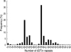
The number of (GT)n repeats ranged from 14-42 and exhibited a bimodal distribution with one peak located at 22 GT repeats and the other at 29 GT repeats (Fig. 1). Because the inducibility of the HO-1 gene promoter was positively correlated with the number of (GT) repeats (19, 20), we divided allelic repeats into 3 subclasses with short, medium, and long (GT)n repeats: short repeats with less than 23 (GT)n were designated as allele class S (short), medium repeats with 23 to 28 (GT)n as allele class M (medium), and long repeats with 29 or more (GT)n as allele class L (long). We had 6 genotypes, S/S, S/M, M/M, S/L, L/M, and L/L, with percentage frequencies (absolute frequencies) of 7.2% (66), 6.9% (63), 3.1% (28), 30.8% (282), 22.7% (208), and 29.4% (269), respectively. The genotypes were categorized into 3 groups: group 1 with S/S genotype, group 2 with S/M and S/L genotypes, and group 3 with M/M, L/M, and L/L genotypes.
Clinical characteristics
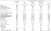
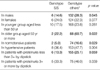
There were no differences in any of the clinical parameters, including medications, among the three genotype groups (Table 1). Group 1 showed no difference in clinical parameters from either of the other two groups. The bilirubin level was not related to the HO-1 genotypes.
The HO-1 genotype and renal impairment of IgA nephropathy at the time of renal biopsy
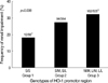
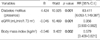
The prevalence of renal impairment differed among the three genotypic groups. In group 1, the renal impairment rate of 18.2% was lower than 32.2% in group 3 (Fig. 2). The univariate factors related to the renal impairment of IgA nephropathy at the time of renal biopsy were age, diabetes mellitus, hypertension, blood pressures, serum cholesterol, serum uric acid, serum albumin, serum bilirubin, hemoglobin, proteinuria 3+ or more by dipstick urine test, and genotype of the HO-1 gene. In multiple logistic regression analysis adjusted for these factors and gender, the HO-1 genotype was an independent factor for renal impairment at diagnosis (Table 2). The risk of renal impairment in patients with single S allele (group 2) was not significantly different from that of group 3 patients but the odds ratio of renal impairment in group 1 was 0.216-fold lower than that in group 3. The odds ratio of impaired renal function in S/S genotype group was 0.249 compared to the other genotypes (group 2 and group 3) and, in S allele group (group 1 and group 2), was 0.598 compared to the other genotypes (group 3).
The HO-1 genotype and renal impairment in subgroups stratified by risk factors
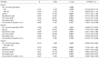
When we stratified patients by gender, age with the criterion of 50 yr, hypertension, and proteinuria 3+ or more by dipstick urine test, which are related to the renal impairment in IgA nephropathy, the rate of renal impairment in group 1 was lower than that in the other groups (group 2 and group 3), especially in subgroups of men, subjects aged 50 yr or more, and normotensive subjects (Table 3). In subjects with proteinuria by dipstick test less than 3+, group 1 tended to have a lower frequency of renal impairment than the other groups, although the difference was not statistically significant. When we stratified the female group by age, the frequency of renal impairment of group 1 was lower than that of the other groups in females aged 50 yr or more (0/3 vs. 44/72, p=0.019).
The risk factors for mortality in IgA nephropathy
There were no deaths among the 64 patients of group 1 whereas there were 11 deaths in the 821 patients of the other groups (p>0.05). All deaths occurred in patients with the L allelic group (11/732). The causes of death were renal failure, nephrotic syndrome, sepsis, heart failure, and subdural hemorrhage. Among univariate risk factors related to mortality, diabetes mellitus, eGFR, and body mass index (BMI) were independent risk factors (Table 4). Three of 24 patients with diabetes mellitus died, as did 8 of the 807 patients without diabetes mellitus (p=0.003). There was no mortality in 624 patients with eGFR 60 mL/min/1.73 m2 or more, 6 deaths out of 196 patients with eGFR 30-59 mL/min/1.73 m2, and 5 deaths among 65 patients with eGFR less than 30 mL/min/1.73 m2 (p<0.001). The subgroups with BMI less than 18.5, 18.5-24.9, 25.0-29.9, and 30.0 kg/m2 or more showed mortality rates of 7.5% (3/40), 0.8% (4/474), 1.0% (2/210), and 0.0% (0/23), respectively. The patients with BMI less than 18.5 kg/m2 showed significantly higher mortality than those with BMI 18.5 kg/m2 or more (p=0.011).
DISCUSSION
In this study, we demonstrated that HO-1 gene promoter polymorphism was related to the prevalence of renal impairment at diagnosis, which is an important risk factor for mortality of IgA nephropathy. HO-1 messenger RNA expression and enzyme activity are greater when the (GT) repeat length in the HO-1 gene promoter region is short but the cut-off point of (GT) repeat number related to HO-1 inducibility repeats of the short allele for HO-1 gene length polymorphism has most commonly been <25 (17, 25, 32, 33), but has also included <28 (24), <27 (21-23), and <23 (19, 20). With the transient-transfection assay with HO-1 gene promoter regions containing various numbers of (GT) repeats, the promoter activity of the HO-1 gene was up-regulated in genes with 16 or 20 (GT) repeats but was not in genes with 29 or 38 (GT) repeats (19). The reporter gene expression driven by the HO-1 gene promoter carrying 22 (GT) repeats was about four- and eight-fold greater than that directed by promoters with 26 and 30 (GT) repeats, respectively (20). Therefore, we grouped the HO-1 gene length polymorphism genotypes into 3 groups with the criteria of less than 23, 23 to 28, and 29 or more (GT) repeats.
The short allele of the HO-1 promoter gene was related to the lower rate of renal impairment defined by the estimated GFR less than 60 mL/min/1.73 m2 as calculated with the modified MDRD equation. Although the MDRD equation was not fully verified to estimate kidney function in the Korean population, we believe that our results are acceptable because the risk of renal impairment defined by the calculated creatinine clearance less than 60 mL/min by the Cockcrauft-Gault equation (34) was also 0.073 (95% confidence interval, 0.014-0.378, p=0.002 by multiple logistic regression analysis) in patients with the S/S genotype compared to the risk in patients with the M/M, L/M, or L/L genotype.
Courtney et al. (25) reported that HO-1 (GT)n promoter polymorphism was not related with the progression to end stage renal disease in patients with IgA nephropathy. In their study, the dependent variable compared between the genotypic groups was the mean age of entry into the renal replacement therapy program. The mean age of entry into this program might be a surrogate marker for renal survival in genetic renal diseases like autosomal dominant polycystic kidney disease but is not usually accepted as a primary outcome in IgA nephropathy. Although the data at the time of renal biopsy might not be the data at the disease onset point, we should consider the clinical status at the time of renal biopsy and adjust the differences of clinical status at diagnosis between patient groups to estimate the risk of disease progression, statistically.
Although the glomerular injury in IgA nephropathy is usually provoked by IgA-induced mesangial cell activation and complement activation leading to pro-inflammatory and pro-fibrotic phenotype transformation in mesangial cells (26), increased oxidative stress was reported to play some roles in the development and progression of IgA nephropathy (27, 28). Chen et al. (28) observed that the renal infiltration of polymorphonuclear leukocytes, which has a high potential for the production of reactive oxygen species, increased in patients with IgA nephropathy. Advanced oxidation protein products increased in progressed IgA nephropathy compared with in stable IgA nephropathy and was an independent risk factor to renal outcome of IgA nephropathy (27).
The HO-1 antioxidant effect is mediated by the removal of pro-oxidant heme, generation of biliverdin and bilirubin, coinduction of ferritin, reduction of hydroxyl radical production, and suppression of the pro-oxidant MCP-1 (reviewed in 1). Renal up-regulation of HO-1 attenuates inducible nitric oxide synthase expression and proteinuria in experimental glomerulonephritis (16). For consideration of the pathogenic mechanism of IgA nephropathy and the roles of the HO-1 gene, the relationship between the renal impairment of IgA nephropathy at diagnosis and the inducibility of the HO-1 gene promoter has logical relevance.
The relationship between HO-1 promoter length polymorphism and renal impairment was evident only in males, partially because of the gender-difference of HO-1 inducibility to various stresses. The induction of HO-1 expression and activity was more enhanced in females than in males after trauma and hemorrhagic shock (35) and myocardial ischemia (36). Actually, the S/S genotype was related to a lower rate of renal impairment in females older than 50 yr, which is older than the mean age of menopause in Korean women of 47 yr (37). The HO-1 inducibility was not related to the renal impairment of patients with more severe clinical findings such as hypertension and high proteinuria, suggesting that the influence of the HO-1 gene might be overwhelmed by the clinical risk factors.
This study had several limitations. The devices for measuring serum creatinine were different among institutions. Jaffe's colorimetric method was used to measure the level of serum creatinine in all of the participating institutions, but the device models differed. We did not standardize the results by calibrating the serum creatinine value of each institution to the result of one standard device. The mortality data were from the national mortality database and we could not assess whether or not the primary cause of mortality was correct.
In conclusion, the short allele of HO-1 gene promoter length polymorphism was related to the lower rate of renal impairment in IgA nephropathy at diagnosis, which is an important risk factor for the mortality of IgA nephropathy.
APPENDIX
Members of The PREMIER group: Institutions that participated in the study (Investigators).
Cheju National University Hospital (Eun Hee Jang), Chonbuk National University Medical School (Won Kim), Chonnam National University Medical School (Nam Ho Kim, Woo Kyun Bae), Chungbuk National University College of Medicine (Hye Young Kim), Chungnam National University College of Medicine (Young-Tai Shin, Kang Wook Lee, Ki-Ryang Na), Daegu Catholic University Medical Center (Ki Sung Ahn), Dankook University Hospital (Jong Tae Cho, Eun Kyeong Lee), Dong-A University College of Medicine (Ki Hyun Kim, WonSuk An, Seong Eun Kim), Ewha Womans University School of Medicine (Choi Gyu Bog, Seung-Jung Kim), Gachon University of Medicine and Science (Woo Kyung Chung, Hyun Hee Lee, Jaeseok Yang, Sejoong Kim), Gyeongsang National University Hospital (Se-Ho Chang), Hallym University College of Medicine (Jung Woo Noh, Young Ki Lee, Seong Gyun Kim, Jieun Oh, Young Rim Song), Inha University College of Medicine (Moon Jae Kim, Seoung Woo Lee), Inje University College of Medicine (Yeong Hoon Kim, Won Do Park), Keimyung university school of medicine (Hyun Chul Kim, Sung Bae Park), Konkuk University School of Medicine (Kyo-Soon Kim), Korea University Anam Hospital (Won Yong Cho, Hyoung Kyu Kim, Sang-Kyung Jo), Korea University Ansan Hospital (Cha Dae Ryong, Kang Young Sun), Korea University College of Medicine Guro Hospital (Young-Joo Kwon), Kyungpook National University School of Medicine (Yong-Lim Kim, Sun-Hee Park, Chan-Duck Kim), Pochon CHA University College of Medicine (Dong Ho Yang), Pusan National University School of Medicine (Ihm Soo Kwak, Soo Bong Lee, Dong Won Lee, Sang Heon Song, Eun Young Seoung), Seoul Medical Center (Su-Jin Yoon), Seoul National University Bundang Hospital (Dong-Wan Chae, Ki Young Na, Ho Jun Chin), Seoul National University College of Medicine Boramae Medical Center (Chun Soo Lim, Yoon Kyu Oh), Seoul National University Hospital (Kook Hwan Oh, Kwon Wook Joo, Yon-Su Kim, Curie Ahn, Jin Suk Han, Suhnggwon Kim), Seoul National University Hosptial Clinical Institute (Hyung Jin Yoon), Sungkyunkwan University School of Medicine (Kyu-Beck Lee), Sungkyunkwan University School of Medicine Samsung Medical Center (Yoon Goo Kim, Jung Eun Lee), Ulsan University College of Medicine, Asan Medical Center (Sang Koo Lee), Yeungnam University College of Medicine (Jun-Young Do, Jong-Won Park, Kyung-Woo Yoon), ordered by alphabet.
 XML Download