

 PDF
PDF Citation
 Print
Print


INTRODUCTION
In Korea, the number of patients with end stage renal disease (ESRD) waiting for transplantation continues to increase (1). Numerous attempts have been made to reduce the number of waiting patients including the donor-exchange program and transplantation with ABO incompatible and/or crossmatch positive donors (2-7). The immune reaction against donor-antigens remains a barrier to the utilization of kidneys from living-donors. Although the proportion of sensitized patients against donor antigens has not been accurately determined to date, 14% of patients on the United Network for Organ Sharing waiting list have a history of a high panel reactive antibody (PRA) titer of more than 80% (8). A preliminary report from our transplant center showed that 15% of the patients on the transplantation waiting list have been considered to be sensitized to human leukocyte antigen (HLA) antigens, and 8% among them have a PRA titer of more than 50% (unpublished data). ESRD patients, with a positive crossmatch donor, have been prevented from proceeding to transplantation because of the potential risk for developing hyperactive rejection resulting in graft failure (9).
MATERIALS AND METHODS
Patients
From August 2006 to January 2008, seven adult sensitized patients with ESRD received living donor kidney transplantation at Seoul National University Hospital and were enrolled in this study. Five patients had a positive crossmatch on the cytotoxicity or flow cytometric assays, and the other two patients had donor specific antibodies (DSAs), despite a negative crossmatch. The patients were scheduled for desensitization pretreatment, followed by living donor kidney transplantation. Both potential recipients and donors were informed of the procedures for a positive crossmatch living-donor transplantation protocol, which was compared with transplantations in cases with a negative crossmatch.
Desensitization protocol
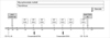
The desensitization protocol used is summarized in Fig. 1. The recipients started taking mycophenolate mofetil (MMF, 750 mg twice daily, p.o.) and tacrolimus (0.05 mg/kg twice daily, p.o., target trough level 10-12 ng/mL) two days before the first plasmapheresis. Methylprednisolone 1,000 mg i.v. was started at the time of the surgery, and the steroid dose was tapered to an oral dose of prednisolone. The combined immunosuppression of the MMF, tacrolimus and prednisolone were continued through the post-transplantation period. In addition, basiliximab (4 mg i.v. on day 0 and day 4) was used for induction therapy in three patients. The plasmapheresis (one plasma volume exchange with 4% albumin and/or fresh frozen plasma) was performed three times a week preoperatively. Intravenous immunoglobulin (IVIG, 100 mg/kg) was administered immediately after each plasmapheresis. After the second and fifth plasmapheresis, crossmatching and DSAs were evaluated. Patients with a negative crossmatch and no donor specific reactivity proceeded to transplantation. The first and second transplant patients received 10 days of OKT3 (muromonab-CD3) (5 mg daily, i.v.) after the transplantation. We modified our protocol to include rituximab instead of OKT3 for the treatment of the third patient through seventh patient. Rituximab (375 mg/m2 of body surface area, i.v.) was administered three days before the first plasmapheresis and one day before transplantation until the CD20 and CD19-positive lymphocytes level was undetectable. The level of CD19-positive lymphocytes was evaluated before the first use of rituximab and two days before transplantation (12).
Assessment of antibody status
Donor T cells were isolated from whole blood for cytotoxicity crossmatching using immunomagnetic beads. A T cell antiglobulin enhanced complement dependent cytotoxicity crossmatch (AHG-CDC) was performed using a serial doubling dilution of the recipient's serum. The last reaction resulting in a >20% cell death, above the background, was considered the anti-donor crossmatch titer. The interpretation of the flow cytometric crossmatch was performed by direct comparison of the fluorescence intensity of the donor T cells after the treatment with the patient's serum to the intensity of donor cells after treatment with a negative control serum. A two-fold change of fluorescence intensity and fluorescence shift was interpreted as positive. The PRA levels were determined by commercial ELISA kit (QID; GTI, Waukesha WI, U.S.A.). Optical density values were assessed to estimate the level of anti-HLA antibodies, using the value recommended by the manufacturer.
Detection and treatment of rejection
All rejection episodes were biopsy-proven. A biopsy was performed in the setting of presumed graft dysfunction manifested by oliguria and an elevation of the serum creatinine. Immunofluorescence staining for IgM, IgA, IgG, C1q, C3, fibrinogen, kappa, lambda, and the complement degradation product of C4d was performed in addition to light microscopic examination of the biopsy samples.
Acute cellular rejection was treated with methylprednisolone pulse therapy (1,000 mg/day for 3 days). Antibody-mediated rejection was treated with plasmapheresis followed by intravenous immunoglobulin (100 mg/kg), rituximab (375 mg/m2 of body surface area, i.v.) plus methylprednisolone pulse therapy. Plasmapheresis/IVIG was delivered daily until DSAs decreased to undetectable levels.
RESULTS
Pre-transplantation characteristics of patients
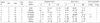
The clinical and serological characteristics of the seven patients are shown in Table 1. Six of the transplant recipients were female. Two patients had received prior transplants, and three patients had the history of blood transfusion. The donors of three patients were biologically unrelated husbands. The average number of donor-recipient HLA mismatches was 2.9, ranging from two to four. Five patients were crossmatch positive with their donors, and three of them had a positive flow cytometric, but negative cytotoxic crossmatch (AHG-CDC). The other two patients had anti-HLA class I/II antibodies with donor specificity even though they did not have a positive crossmatch by cytotoxicity or flow cytometric analysis.
Negative conversion of positive crossmatch
All seven patients achieved a negative crossmatch and/or negative conversion of DSAs at the time of transplantation. As crossmatching was not performed after each plasmapheresis, the required number of sessions of plasmapheresis/IVIG to achieve a negative conversion could not be estimated accurately. All patients had an acceptable alloimmunization status after five plasmapheresis/IVIG treatments. Some patients were given several additional sessions of pre-/post-transplantation plasmapheresis after the crossmatch became negative.
Graft survival and renal function
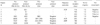
All patients are alive with normal renal function at a mean follow-up of 13.2±7.8 months. The post-transplantation status and outcomes are shown in Table 2. In patient 6, the serum creatinine increased up to 1.9 mg/dL 62 days after the transplantation, which appeared to be associated with tacrolimus toxicity; the serum creatinine level decreased to 1.1 mg/dL after reduction of the tacrolimus dose. The mean serum creatinine level at the last follow-up was 1.1±0.1 mg/dL.
Acute rejection
Acute cellular rejection occurred in two patients, and antibody-mediated rejection occurred in one patient. Patients 1 and 5 had a renal biopsy 21 days and 13 days after transplantation, respectively. At the time of the renal biopsy the serum creatinine level was 1.3 mg/dL in both patients. Although the renal biopsy revealed acute cellular rejection with tubulitis and interstitial infiltration of inflammatory cells, these episodes were subclinical without any definite evidence of allograft dysfunction except for mild elevation of the serum creatinine. The patients were treated with bolus methylprednisolone (1,000 mg/day for 3 days). Patient 6, who received a renal transplant from her brother, was enrolled in the desensitization protocol because of the pre-protocol presence of DSAs (A24, DR15). The DSAs were eliminated after two plasmapheresis/IVIG treatments, and the renal transplantation was performed after a total of five treatments. Since the serum creatinine was persistently elevated on three consecutive measurements (peak level of 1.7 mg/dL), a renal biopsy was performed on post-operative day 14. The renal biopsy showed antibody-mediated rejection with peritubular capillary C4d staining. The donor specific antibody (DR15) was detected again at the time of the biopsy. The patient was treated with bolus methylprednisolone (1,000 mg/day for 3 days) followed by a tapering dose of prednisolone to the previous dose, and plasmapheresis followed by IVIG (100 mg/kg) for five days. The serum creatinine was 1.1 mg/dL at hospital discharge.
Complications
No adverse events associated with infusion of OKT3, rituximab or IVIG were observed. Bleeding occurred in four patients; three patients received packed-RBC transfusions, and one patient underwent surgical exploration for bleeding control. A wound infection occurred in one patient, but it was controlled with intravenous antibiotics.
DISCUSSION
Although kidney transplantation in sensitized patients might be a barrier for successful transplantation, the recent advances in desensitization enabled the clinicians to expect good outcomes in graft survival as well as in patient survival (10-13). Here, we described successful kidney transplantation in seven recipients who were previously sensitized.
The characterization of anti-donor antibodies is a major issue in transplantation of sensitized patients (14-16). Anti-donor antibodies can be detected by several techniques that vary with respect to their sensitivity and specificity. Many previous studies enrolled patients with positive cytotoxicity determined by AHG-CDC. However, recent report has identified patients with a positive pre-transplant T-flow despite a negative T-cytotoxicity who have developed acute rejection associated with impaired long-term graft survival (17). The role of the DSAs in patients with a negative crossmatch is controversial (18, 19). One patient in our case series, who enrolled in the desensitization protocol, due to the pre-transplant presence of DSAs (A24, DR15) despite a negative T-cytotoxicity and a negative T-flow, developed antibody-mediated rejection 14 days after transplantation. The DSAs were eliminated after two plasmapheresis/IVIG treatments; however, they were detected again at the time of rejection. The culprit antibody was anti-DR15. Large long-term studies are needed to evaluate the clinical influence of donor specific antibodies on long-term graft survival.
The proposed protocols that we used were different from prior procedures in several aspects including the recruitment of eligible patients for desensitization, use of rituximab or anti-thymocyte globulin (ATG)/OKT3 and the protocol for obtaining renal biopsies. IVIG plays an important role in desensitization protocol (10-13). There are two IVIG protocols: high-dose IVIG and plaspmapheresis/low-dose IVIG (PP/IVIG). Each protocol has advantages and disadvantages. High-dose protocol is less expensive, easy to administer, and does not need live donor. But, some patients might not respond to high-dose IVIG, and antibody removal might be less rapid. Low-dose IVIG could remove DSAs more rapidly and effectively, although it needs live donor, more costs and resources. We have selected the PP/IVIG protocol because we believe it provides more reliable elimination of DSAs in the setting of living donor kidney transplantation. Although PP/IVIG can cause complications such as infections and immune complex disease, short course therapy is considered safe. Our results demonstrated that plasmapheresis/IVIG was effective for decreasing the PRA as well as donor specific antibodies (Table 2).
Rituximab may be used as part of a preconditioning regimen for sensitized patients. It could effectively diminish the post-plasmapheresis rebound and post-transplantation production of anti-donor antibodies by depleting B cells. Though many desensitization protocols including rituximab have been introduced, the dosing schedule of rituximab has not been established. Montgomery et al. (12) reported the weekly regimen of rituximab; they administered rituximab 1 month prior to PP/IVIG and dosed (375 mg/m2 body surface area per week, not to exceed four total dose) until the CD20 and CD19-positive lymphocytes level was undetectable. Gloor et al. (11) introduced single-dose protocol; they administrerd rituximab (375 mg/m2 body surface area) 4 days before transplantation. However, recent report demonstrated failure of rituximab therapy in crossmatch positive renal transplantation (21). Modifying weekly regimen, we administered rituximab (375 mg/m2 body surface area) at D-19 and D-1 until the CD20 and CD19-positive cells were undetectable.
MMF was also used for inhibition of B cell proliferation. Although successful protocols without rituximab or splenectomy have been reported (10, 21), rituximab appears to be beneficial for positive crossmatch transplantation. The use of rituximab has been associated with false positive cytotoxic and flow cytometric results. Although pronase treatment may overcome this interference (22), it was not used in this study. Fortunately, none of the five patients that received rituximab had a false-positive-crossmatch after the use of rituximab. An improved solid-phase assay might be included in future desensitization protocols. OKT3 and antithymocyte globulin have been shown to be effective in crossmatch positive transplantations (10). After the second patient, we did not include OKT3 in our protocol. Although two of the five patients without OKT3 experienced acute cellular rejection, the actual contribution made by OKT3 to the outcomes in the present study could not be determined.
The graft survival in the present study compared favorably with the results reported by previous studies on positive crossmatch transplantation (10-13). Generally, our protocol consisted of an intensive regimen. Serious complications occurred in only two patients. One required an exploratory laparotomy for bleeding control, and the other received antibiotics intravenously for the control of a wound infection.
In conclusion, sensitization is not necessarily a contraindication to the living donor kidney transplantation. Using a protocol including plasmapheresis/IVIG, tacrolimus, MMF, and rituximab, seven sensitized patients had successful kidney transplantation. Such protocols for desensitization as well as donor exchange programs would expand the donor pool, and may contribute to resolving the organ shortage in transplantation.
 XML Download