

 PDF
PDF ePub
ePub Citation
 Print
Print


INTRODUCTION
Endometrial stromal sarcoma (ESS) is a rare neoplasm comprising only 0.2% of all uterine malignancies and usually occurs as a primary tumor of the uterine corpus (1).
ESS of extrauterine origin is a very rare type of neoplasm and arises in sites, such as the ovary, the pelvic cavity, mesentery, omentum, and serosal or intramural portion of the large intestine (2-4). The tumor is usually classified into two subtypes, low-grade ESSs and undifferentiated sarcomas with the latter subtype carrying a very poor prognosis (5, 6).
We describe a 60-yr-old woman with low-grade ESS presented as prevesical mass mimicking urachal mass.
CASE REPORT
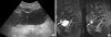
A 60-yr-old woman presented with incidentally detected prevesical mass. Her history was remarkable in that she had a total hysterectomy by lower median abdominal incision for endometriosis 11 yr ago. The family history was unremarkable. Physical examination showed no palpable abdominal mass. Routine blood tests, blood chemistry and urine test revealed no abnormal findings. In the previous ultrasonographic examination, heterogeneous hyperechoic pelvic mass was incidentally detected, and she was transferred to our hospital on the impression of urachal tumor (Fig. 1A).
Magnetic resonance imaging of the abdomen and pelvis showed a mass located anterior to the bladder in the prevesical space of Retzius and helped to establish the relation of the tumor to the bladder in the sagittal plane (Fig. 1B).
Complete excision with bladder sparing was successful by lower median abdominal incision. At surgery, two tumor masses were founded in the prevesical space of retzius and had not infiltrated the wall of the bladder and urachus.
Grossly, well-circumscribed yellowish tumors were 4.5×2.5×2 cm, 1.5×1×1 cm in size, respectively. Microscopically, the lesion was characterized infiltrating nest of tumor cells. The tumor consisted of relatively large cells resembling those of the endometrial stroma, which encircled small vessels (Fig. 2A). Tumor cell nuclei was round to ovoid and a small amount of cytoplasm was present. Mitotic activity was less than 10 mitotic figures per 10 high power field (HPF) (Fig. 2B). Immunohistochemically, the tumor cells were positive for CD10 but negative for cytokeratin. Most of the cells were also negative for estrogen receptor, and positive for progesterone receptor (data not shown). Based on these findings, a low-grade ESS was diagnosed. Her postoperative recovery was uneventful. She was free of symptoms at the last follow-up, 15 months after the abdominal surgery, with no evidence of recurrence.
DISCUSSION
ESS usually occurs as a primary tumor of the uterine corpus and is rare neoplasm. ESS of extrauterine origin is very rare. Because of the rarity of ESS, very little is known about its pathogenesis. However, there have been some reports in which ESS is considered to be associated with endometriosis (2-4). It arises in area of pre-existing endometriosis and also arises in cases that are not associated with endometriosis.
These are explained by two hypotheses. One is malignant transformation of endometriosis. Various types of neoplasm have been observed in patients with endometriosis.
Histologically, endometrial adenocarcinoma is the most (about 70%), and sarcoma is the second common malignancy (12%) (7). But exact incidence of ESS in endometriosis is not well described. Heaps et al. (7) reported that malignant tumor arising in endometriosis involved ovary (78.7%) and extragonadal site (21.3%).
Majority of extragonadal sites were rectovaginal septum (4.3%), colon (4.3%), vagina (2%), and pelvic peritoneum (5.7%). The other theory is a de novo tumor, potentially derived from submesothelial pluripotential Mullerian cells. The pluripotential Mullerian epithelium is considered to be widely distributed in the abdominal and pelvic cavities (4).
In the present case, the patient had undergone total hysterectomy for endometriosis 11 yr ago, and the tumor was localized in the prevesical space. So, we assumed that it arose from prevesical endometriosis because the patient had a history of endometriosis but there is no direct evidence.
Previously, the tumor is usually classified into two categories, low and high-grade, based on differences in mitotic activity, with the latter subtype carrying a very poor prognosis (5). Recently, the World Health Organization classification for tumors of the uterus classifies the tumors based primarily on tumor margin status and cytologic features (6). Low-grade ESSs are clinically indolent malignancies with minimal cytologic atypia and proliferative activity. Undifferentiated sarcomas (previously categorized as high-grade endometrial stromal sarcomas) are highly aggressive malignancies that show substantial cytologic atypia and high mitotic activity.
Young et al. (3) reported that the mitotic rate of the extrauterine ESS correlated with the prognosis but Chang et al. (2) reported that neither mitotic index nor cytologic atypia in primary extrauterine ESS were predictive of tumor recurrence or death from tumor. In their series, 77% of patients whose tumors had a mitotic index of less than 10 per HPF had one or more recurrences and 30% died from their neoplasm. The behavior of the primary extrauterine ESS was more reminiscent of high-grade primary uterine ESS than low-grade primary uterine ESS (2). Therefore, close observation and extended follow-up will be required in extrauterine ESS, regardless of mitotic activity.
Low-grade ESS has slow growth and indolent course with a tendency for late recurrence; approximately 50% of cases of low-grade ESS develop recurrent disease (6). A relatively long-term survival, despite tumor dissemination, has been reported in low-grade ESS cases. Both recurrent and metastatic lowgrade sarcomas can be successfully treated with surgical excision, radiation, progestin therapy, or a combination therapy (6). Considering highly recurrent nature of extrauterine ESS or low-grade ESS, a life long follow-up is necessary for this patient.
Tumor localized in the prevesical space is very rare but urachal tumor has been most commonly presented in prevesical space. However, when patient with endometriosis presented with prevesical mass in Retzius space, endometriosis and its possible malignant changes should be taken into account in
differential diagnosis.

 XML Download