

 PDF
PDF ePub
ePub Citation
 Print
Print


INTRODUCTION
Genomic DNA hypomethylation has been observed in the peripheral blood mononuclear cells of leukemia patients and in tumor cells of patients with B-cell lymphoproliferative diseases (1). DNA methylation may affect karyotypic stability, may influence euchromatin-heterochromatin interactions, and has been correlated with disease progression (2). On the other hand, promoters of some tumor suppressive genes are often hypermethylated (3-6). For example, some patients with lymphomas do not express tumor suppressor genes because the promoters of these genes are methylated.
Leukemia is a hematological cancer of the bone marrow and lymphatic system. In leukemia, bone marrow produces a large number of abnormal white blood cells, which overwhelm the other types of blood cells, including red blood cells and platelets, thus impairing the production of normal white blood cells. In clinical classification, leukemia can be classified as acute myeloid leukemia (AML) and chronic myeloid leukemia (CML). AML is a serious and lethal disease that affects adults. CML is a clonal disease of stem cell origin that is characterized by the presence of the Philadelphia chromosome (Ph+), which has been named t(9,22)(q34:q11). Its fusion gene product, Bcr-Abl, is a constitutively active tyrosine kinase. These facts suggest that somewhat different mechanisms may be involved in AML and CML. Nevertheless, the differential factors through which they produce a different type of leukemia are not yet completely understood.
In hematopoietic malignancies, hypermethylation of several genes including E-cadherin, DAP kinase, estrogen receptor (ER) alpha, and p15INK4B are associated with gene inactivation (7-10). Genes, such as DAB2IP, DLC-1, H-cadherin, ID4, Integrin α4, RUNX3, SFRP1, and SHP1, has been identified as being implicated in aberrant DNA methylation during development of human malignancy (11-18). In order to gain insight into the differential epigenetic alterations in leukemia, we investigated the methylation statuses at selected locus of these genes in AML and CML patients using a methylation-specific polymerase chain reaction (MSP).
MATERIALS AND METHODS
Sample collection
Diagnostic bone marrow samples were obtained from 23 patients with AML and 21 patients with CML. The samples were gathered by the Division of Hematology/Oncology (Department of Internal Medicine, Korea University Medical Center, Seoul, Republic of Korea) and analyzed by pathologist. Institutional review board approval and informed consent were obtained (KUMC-IRB-2006011-P-1, KUMCIRB-2006012-P-2). Of the AML patients, 17 males and 6 females were included, with ages ranging from 26 to 78 yr, at a median age of 45.61 (SD, 15.56) yr. Of the CML patients, 11 males and 10 females were included, with ages ranging from 18 to 75 yr (mean±SD, 49.91±16.53). As controls, 22 normal peripheral bloods were obtained from healthy volunteers (11 males and 10 females) ranging from 20 to 78 yr of age (mean±SD, 45.36±20.64).
Sodium bisulfite treatment and MSP
Chemical modification was performed as described previously, with minor modifications (19). In brief, 1 µg of genomic DNA was denatured by incubation with 0.2 M NaOH for 10 min at 37℃, followed by the addition of 550 µL of 3 M sodium bisulfite (pH 5.0) (Sigma, St. Louis, MO, U.S.A.) and 10 mM hydroquinone (Sigma), which was brought to a final volume of 600 µL. The mixtures were incubated at 55℃ for 16 hr, and the modified DNA was then desalted with the Wizard Clean-Up system (Promega Corp., Madison, WI, U.S.A.). We performed polymerase chain reaction (PCR) using specific PCR primers capable of distinguishing between methylated and unmethylated DNA sequences. The primers for the unmethylated and methylated DNA sequences, PCR product size, and annealing temperature are shown in Table 1. The MSP primer sets were selected at the 5'-CpG island regions of genes using the MethPrimer software (www.urogene.org). The PCR conditions were as follows: initial denaturation and hot start at 95℃ for 5 min, and cycles consisting of 30 sec at 95℃, 30 sec at the annealing temperature, and 30 sec at 72℃. In addition, SCK, a human cholangiocarcinoma cell line, was used as positive control for Integrin α4 gene (20).
Statistical analysis
MSP results were analyzed as a dichotomous variable based on the presence or absence of gene methylation. The MSP results of tumor and normal samples were compared and analyzed with the Pearson chi-square test (Version 12; SPSS Inc., Chicago, IL, U.S.A.). Statistical significance was defined as a P value of <0.05.
RESULTS
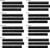
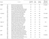
The multiple genes were found to be methylated in bone marrow from patients with AML or CML. Specifically, the frequencies of promoter hypermethylation at selected locus in the 23 AML samples were: 78.3% (18/23) for SHP1, 65.2% (15/23) for ID4 and SFRP1, 26.1% (6/23) for H-cadherin, 8.7% (2/23) for DLC-1, and 4.3% (1/23) for DAB2IP and RUNX3. The frequencies of DNA hypermethylation at selected locus in the 21 CML samples were: 28.6% (6/21) for SHP1, 19.0% (4/21) for H-Cadherin, 14.3% (3/21) for ID4, 9.5% for (2/21) for SFRP1, and 0% (0/21) for DAB2IP, DLC-1, Integrin α4, and RUNX3 (Fig. 1). However, promoter hypermethylation of the 22 normal peripheral bloods was observed less frequently (Table 2).
There was a statistically significant difference between normal peripheral bloods and AML with respect to the frequencies of methylation of ID4, SFRP1, and SHP1 (Pearson chi-square test; P<0.0001, P<0.0001, and P<0.0001, respectively) and between normal peripheral bloods and CML with respect to the frequencies of SHP1 methylation (Pearson chi-square test; P=0.007). Furthermore, there was a statistically significant difference between the DNA methylation frequencies of AML patients and CML patients. The frequencies of DNA methylation of ID4, SFRP1, and SHP1 were higher in AML compared to those in CML (P=0.001, P<0.0001, and P=0.001, respectively) (Table 2). In contrast, no statistical differences between AML and CML were detected in other genes such as DLC-1, DAB2IP, H-cadherin, Integrin α4, and RUNX3. Promoter methylation results at selected locus of eight genes with MSP method were shown in Fig. 1. These results suggest that there may be differential epigenetic modification between AML and CML.
DISCUSSION
Differential epigenetic alteration is observed in AML and CML patients. The results of the present study demonstrate the substantially increased frequency of promoter hypermethylation in ID4, SFRP1, and SHP1 genes in AML compared to CML.
Leukemia, a heterogeneous group of hematopoietic malignancies that occur worldwide, includes acute and chronic myeloid leukemia. Despite many important advances in understanding the different biological and cytogenetic aspects of acute and chronic leukemia, a number of questions remained unanswered. It is understood that some kinds of leukemia present specific cytogenetic alterations (21). Different types of leukemia usually have specific epigenetic modifications that cause the activation of oncogenes and, in particular, the formation of abnormal fusion genes such as AML1-ETO (22, 23), a fusion protein resulting from t(8,21) translocation that commonly occurs in AML. Another typical example is the fact that Bcr-Abl is a constitutively active, cytoplasmic tyrosine kinase that is generated by t(9;22) translocation in more than 95% of CML (24).
The central role of epigenetic modification of these genes in leukemia development promoted them as reasonable targets for understanding the differential mechanism between acute and chronic leukemia. In the present study, ID4, SFRP1, and SHIP1 were hypermethylated in AML compared to CML. These results suggest that the epigenetic modification of these genes may play specific roles in the development of AML rather than CML and, thus, promoted them as ideal targets for drug development to treat AML. In the future, it is clearly imperative to understand the patho-physiological differential mechanisms of these genes in AML, and to finally explore novel therapeutic strategies.
In present study, methylation frequencies of SHP1 gene were the high detected in CML as well as AML. SHP1 is a member of the SHP family of proteins, cytoplasmic protein tyrosine phosphatase (PTP), and has been known as a candidate tumor suppressor gene in lymphoma, leukemia and other cancers (25). Also, the hypermethylation of SHP1 gene was frequently detected in several human cancers and the reduced expression of the SHP1 gene in various types of leukemias and lymphomas mainly occurred by promoter methylation (26-28). Therefore, these results indicate that aberrant DNA methylation of SHP1 gene may be related to the tumorigenicity of myeloid leukemia.
The methylation frequencies of the DAB2IP, DLC-1, and RUNX3 genes were detected at different levels in acute and chronic leukemia patients; however, the difference was negligible. In some genes like Integrin α4 gene, the promoter methylation was not observed in most of the cases. However, hypermethylation of these genes was frequent event in esophageal squamous cell carcinomas, lung cancers, and gastric cancers (11, 12, 15, 16). Thus, DNA methylation of these genes may be not related to tumorigenicity of AML/CML in contrast with other cancers.
In summary, we have identified that aberrant DNA methylation of SHP1 is a frequent event in AML and CML. Also, the frequencies of DNA methylation of several methylation-controlled genes, including ID4, SFRP1, and SHP1, were higher in AML patients compared to those in CML patients. Although these results should be confirmed, more comprehensive studies are necessary involving various known risk factors such as smoking and occupational carcinogens, and genetic susceptibilities. Our results suggest that aberrant DNA methylation of SHP1 may be related to the tumorigenicity of AML and CML and hypermethylation of ID4, SFRP1, and SHP1 genes may contribute to the pathogenesis mechanism of AML specifically.

 XML Download