

 PDF
PDF ePub
ePub Citation
 Print
Print


INTRODUCTION
Neuroblastoma is the most common extracranial solid tumor in children. The most important clinical and biologic prognostic factors currently used to determine treatment are the age of the patient at diagnosis, stage of disease, N-myc amplification status, DNA ploidy and Shimada histology (1-10). Serum markers such as lactate dehydrogenase (LDH), ferritin and neuron-specific enolase (NSE) also may be predictive of survival (11-13).
Neuroblastoma may develop at any site of sympathetic nervous system tissue. Approximately 80% of all neuroblastomas occur within the abdomen (14-16), while other neuroblastomas grow in the mediastinum, at cervical and other rare sites. Tumors originating from different sites might have different clinical and biological characteristics. Indeed, the tumor site has been proposed to be prognostically important in a few studies (14-22). For example, mediastinal tumors have been reported to be associated with a more favorable course than are abdominal tumors (14-18). However, there is limited information on the prognostic significance of tumors originating from sites other than the abdomen and thorax. Furthermore, a direct comparison between extra-abdominal neuroblastomas, including mediastinal tumors, and abdominal neuroblastomas is rare in the medical literature. Therefore, in the present study, the clinical and biological prognostic characteristics of extra-abdominal neuroblastomas compared to abdominal neuroblastomas were investigated.
MATERIALS AND METHODS
Patients
From February 1997 to December 2007, all patients who were newly diagnosed with neuroblastoma at the Samsung Medical Center were retrospectively reviewed for the present study. A diagnosis of neuroblastoma was made based on either histological examination of the tumor specimens or bone marrow infiltration by neuroblastoma cells and elevated urine catecholamine levels. The N-myc copy number was determined using Southern blot analysis or the quantitative reverse transcriptase-polymerase chain reaction. The patients were staged according to the international staging system (INSS) reported by Brodeur et al. (23) and the extent of the disease was evaluated using computerized tomography, a technetium-99 bone scan, bilateral bone marrow aspirates and biopsy specimens and an iodine-131 or 123-metaiodobenzylguanidine scan. Measurement of serum LDH, ferritin, NSE and urine vanillylmandelic acid (VMA) was part of a routine evaluation at diagnosis.
Treatment
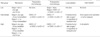
Table 1 summarizes the treatment plans. Patients were treated according to age-, stage- and N-myc-related protocols. Briefly, for patients with low-risk tumors, the primary treatment was surgery with (stage 2) or without (stage 1) postoperative chemotherapy. For patients with intermediate-risk tumors, preoperative chemotherapy followed by surgery and postoperative chemotherapy was the treatment regimen. While local radiation therapy to the primary site was given only if gross total tumor removal was not possible in the early period of the study (diagnosis until December 2003), all patients received local radiation therapy regardless of resectability later in the study (diagnosis from January 2004). High-dose chemotherapy and autologous stem cell rescue (HDCT/ASCR) was provided when gross tumor persisted despite the above-mentioned conventional therapies. For patients with high-risk tumor, HDCT/ASCR was routinely given as consolidation therapy after preoperative chemotherapy, surgery and postoperative chemotherapy (24). Thirteen-cis-retinoic acid was usually administered for 10 cycles (125 mg/m2/day for 14 days per every 4 weeks) to treat possible residual tumor cells for intermediate- and high-risk patients after completion of the scheduled chemotherapy. For high-risk patients, immunotherapy using interleukin-2 was given (2×10⁶ U/m2/day, subcutaneous injection for 5 consecutive days per every 4 weeks) with or without preceding induction therapy (3×10⁶ U/m2/day, continuous infusion for 5 consecutive days per week for 2 weeks) after HDCT/ASCR. IL-2 immunotherapy was usually initiated when the platelet count exceeded 50×109/L without a transfusion after HDCT/ASCR. The first dose of IL-2 was begun on the first day of the 13-cis-retinoic acid treatment cycle and was applied until 1 yr after HDCT/ASCR (24).
Statistics
The clinical and biological characteristics were compared according to the site of tumor origin (extra-abdominal versus abdominal). Regarding the clinical prognostic factors, age, sex and INSS stage were compared between the two groups. For the biological prognostic factors, the Shimada classification, N-myc amplification (≥3 copies), serum LDH, ferritin, NSE and urine VMA were compared between the two groups. Differences in the frequencies of the clinical and biological factors were compared using the Pearson chi-square test. Differences in the levels of tumor markers were compared using the Mann-Whitney U test. Levels of tumor markers are presented as the median values along with the ranges. Overall survival (OS) and event-free survival (EFS) rates along with 95% confidence interval (CI) were estimated using the Kaplan-Meier method. An event was defined as the occurrence of relapse/progression or treatment-related death. The differences in the survival rates between the two groups according to the clinical and biological factors were compared using the log-rank test. Statistical significance was accepted when the P values were less than 0.05.
RESULTS
Clinical and biological characteristics of all patients at diagnosis
From February 1997 to December 2007, 143 consecutive patients were newly diagnosed with neuroblastoma. Among 143 neuroblastomas, 115 (80.4%) tumors originated from the abdomen, 26 (18.2%) from extra-abdominal sites (thorax in 21, neck in 4 and orbit in 1) and two (1.4%) from unknown primary sites. Table 2 lists the clinical and biological characteristics of the 141 patients, except for the two patients with tumors from unknown primary sites. The median age at diagnosis of the 141 patients (81 boys and 60 girls) was 25 months (range 1-129), and 101 (71.6%) patients were over 12 months of age at diagnosis. Sixty-three (44.7%) tumors were localized (stage 1 in 16 patients, stage 2 in 12 and stage 3 in 35) and 78 (55.3%) tumors were disseminated (stage 4 in 77 patients and stage 4S in 1). Fifty-one (37.8%) of 135 tumors analyzed were N-myc amplified (≥3 copies), and 72 (53.7%) of 134 tumors analyzed had unfavorable Shimada pathology. The number of low-, intermediate- and high-risk patients were 28 (19.9%), 32 (22.7%), and 81 (57.4%), respectively. One hundred-eight, 120, 133, and 132 patients had serum LDH, ferritin, NSE and 24-hr urine VMA measured preoperatively at diagnosis. The values measured postoperatively were not included in the analysis. The levels of the serum LDH, ferritin, NSE and 24-hr urine VMA were a median of 1,352 (range 176-9,944) IU/L, 142 (10-6,690) ng/mL, 68 (7-1,081) ng/mL and 5.6 (range 0.1-101.8) mg/day, respectively.
Treatment
All 16 patients with stage 1 disease received surgery only and all 12 patients with stage 2 disease received surgery followed by postoperative chemotherapy. Among 32 intermediate-risk patients, 10 received single or double HDCT/ASCR because of persistent gross tumor or tumor relapse. All 81 high-risk patients except five patients (progression in 2 patients, early death in 2 and refusal of HDCT/ASCR in 1) received single or double HDCT/ASCR, or are scheduled to receive HDCT/ASCR.
Clinical and biological characteristics with respect to the site of tumor origin
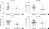
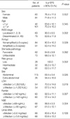
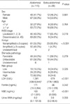
Table 3 shows the clinical and biological characteristics of the extra-abdominal and abdominal neuroblastomas. As for sex, there was no difference between the two groups. The median age at diagnosis in the abdominal and extra-abdominal group was 28 (range 1-129) months and 22.5 (range 7-74) months, respectively (P=0.484). In addition, there was no difference in the number of patients older than 1 yr of age at diagnosis between the two groups. The frequency of disseminated tumors (stage 4 and 4S) was lower in the extra-abdominal group than in the abdominal group (34.6% vs. 60.0%, P=0.019). The frequency of N-myc amplified tumors was also lower in the extra-abdominal group than in the abdominal group (4.2% vs. 45.0%, P<0.001). Accordingly, the proportion of high-risk tumors was lower in the extra-abdominal group than in the abdominal group (34.6% vs. 62.6%, P=0.026). Furthermore, the frequency of unfavorable Shimada pathology was lower in the extra-abdominal group than in the abdominal group, albeit this difference was not significant. The levels of serum LDH, ferritin, NSE and urine VMA were lower in the extra-abdominal group than in the abdominal group (median 679 vs. 1,391 IU/L, P<0.001, Fig. 1A; median 36 vs. 171 ng/mL, P=0.042, Fig. 1B; median 17 vs. 103 ng/mL, P<0.001, Fig. 1C; median 3.9 vs. 6.2 mg/day, P=0.056, Fig. 1D). The characteristics of the five extra-thoracic and extra-abdominal tumors were very similar to those of thoracic tumors. All five tumors were localized (stage 1 in 1 patient, stage 2 in 2 and stage 3 in 2). All of four both extra-thoracic and extra-abdominal tumors analyzed had non-amplified N-myc results. The levels of serum LDH, ferritin and NSE were very low (median 504 IU/L, 24 ng/mL and 12 ng/mL, respectively).
Treatment outcome with respect to the site of tumor origin
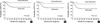
Tumors relapsed or progressed in 16 patients and treatment-related mortality occurred in 14 patients. Therefore, 111 out of 141 patients remained event free with a median follow-up of 47 months (range 1-131) from diagnosis. The probability of 5-yr OS and EFS rate (±95% CI) for all 141 patients was 76.3±8.5% and 73.9±8.1%, respectively. The probability of 5-yr EFS rates and the results of the unstratified log-rank test with respect to the clinical and biological characteristics are listed in Table 2. The probability of 5-yr EFS rate in patients with extra-abdominal tumors was higher than in patients with abdominal tumors (94.4±10.6% vs. 69.4±9.4%, P=0.026). Fig. 2A shows the EFS curves for patients with extra-abdominal and abdominal tumors. All but one patient in the extra-abdominal group are currently event free. When the analysis was confined to only the patients older than 1 yr of age at diagnosis, the probability of 5-yr EFS rate was higher in patients with extra-abdominal tumors than in those with abdominal tumors (92.3±14.5% vs. 65.3±11.7%, P=0.047, Fig. 2B). Similarly, when the analysis was confined to only the patients with stage 4 tumors, the probability of 5-yr EFS rate was higher in patients with extra-abdominal tumors than in those with abdominal tumors (87.5±22.9% vs. 59.2±12.8%, P=0.133, Fig. 2C), albeit this difference was not significant. All five patients with both extra-thoracic and extra-abdominal tumors remained event free with a median follow-up of 123 months (range 10-131) from diagnosis.
DISCUSSION
Neuroblastoma may develop at any site of sympathetic nervous system tissue. Tumors originating from different sties might have different clinical and biological characteristics. Indeed, a few studies have reported that the tumor site was prognostically important in neuroblastoma (14-22). However, most studies addressing the prognostic significance of the tumor site of origin compared thoracic neuroblastomas and others, and found that thoracic neuroblastomas were associated with more favorable clinical features than neuroblastomas originating from other sites, including the abdomen (14-18). The investigators suggested that the clinical features of rare neuroblastomas, originating from both extra-thoracic and extra-abdominal sites, might be similar with those of thoracic neuroblastomas. Therefore, the present study was designed to determine the differences in the clinical and biological features between extra-abdominal and abdominal neuroblastomas.
We have shown that the biology of extra-abdominal neuroblastomas is indeed different from that of abdominal neuroblastomas. The results of the present study revealed that the extra-abdominal neuroblastomas were associated with more favorable prognostic factors than were the abdominal neuroblastomas. Frequencies of disseminated tumors, N-myc amplified tumors and tumors with unfavorable Shimada pathology were lower in patients with extra-abdominal tumors compared to those with abdominal tumors. In addition, levels of the serum LDH, ferritin, NSE and urine VMA were lower in the extra-abdominal tumors than in the abdominal tumors. When the analysis was confined to only the patients older than 1 yr of age at diagnosis, patients with stage 4 tumors or with high-risk tumors, the levels of LDH and NSE were significantly lower in patients with extra-abdominal tumors compared to those with abdominal tumors (data not shown). The characteristics of both extra-thoracic and extra-abdominal tumors were very similar to those of thoracic tumors. In addition, the results of this study demonstrated that patients with extra-abdominal neuroblastomas had a more favorable long-term outcome than did the patients with abdominal neuroblastomas. All patients with both extra-thoracic and extra-abdominal tumors are currently event free.
It is not clear whether the more favorable outcome with extra-abdominal tumors could be attributed to their association with favorable clinical and biological features, or whether the extra-abdominal site is an independent favorable prognostic factor; this is because a multivariate Cox analysis was not possible in the present study because of the small number of patients. However, the long-term outcome was better in the patients with extra-abdominal tumors than in patients with abdominal tumors even when the data was corrected for age at diagnosis, stage or risk-group.
Prior studies on thoracic neuroblastomas have reported a female prevalence (2, 15); however, there was no female prevalence in the extra-abdominal group in the present study. When the analysis was confined to only the patients with thoracic neuroblastomas, there was no female prevalence. According to the study reported by Morris et al. (14), patients with thoracic neuroblastomas were younger than patients with other sites of origin. However, there was no difference in the age at diagnosis between the extra-abdominal and abdominal neuroblastomas in the present study. In addition, when the analysis was confined to 21 patients with thoracic neuroblastomas, there was no difference in the age at diagnosis when compared with other neuroblastomas. In the report by Suita et al. (15), there was also no difference in age between thoracic and nonthoracic patients. Adams et al. (16) confirmed that children with thoracic neuroblastomas have a more favorable outcome, and this was not because of earlier clinical presentation or earlier expression of symptoms.
During the last decade, HDCT/ASCR has improved the survival of patients with high-risk neuroblastoma (24-29). Most patients with high-risk tumors in the present study received tandem HDCT/ASCR and the probability of 5-yr EFS among the high-risk patients was over 60%. However, despite the remarkable improvement in the survival rate in the present study compared to previous studies addressing the prognostic significance of tumor site in neuroblastoma, there is still a significant difference in survival between patients with extra-abdominal neuroblastomas and those with an abdominal origin. This finding suggests that the tumor site of origin is a prognostic factor even with the most current treatment protocols.
This is the first study to investigate the differences in the clinical and biological characteristics between extra-abdominal and abdominal neuroblastomas. The results of the present study show that neuroblastomas originating from extra-abdominal sites are associated with better clinical and biological characteristics and they are associated with a better outcome than those originating from the abdomen. Further study is needed to explain these findings and to determine whether an extra-abdominal site is an independent favorable prognostic factor.
 XML Download