

 PDF
PDF ePub
ePub Citation
 Print
Print


INTRODUCTION
The incidence of pulmonary disease caused by nontuberculous mycobacteria (NTM) in HIV-negative patients is increasing worldwide (1-3). A substantial proportion of these patients has no predisposing risk factors, such as pre-existing lung disease or demonstrable immunodeficiency. These patients are predominantly elderly women with no history of smoking who complain of chronic cough and sputum (1-3). Chest high-resolution computed tomography (HRCT) scans reveal characteristic findings of bilateral multifocal bronchiolitis (well-defined small nodules and branching centrilobular nodules, or tree-in-bud pattern) and bronchiectasis (4-9). Mycobacterium avium complex (MAC) and Mycobacterium abscessus are well-recognized organisms that cause these forms of NTM lung disease (4-9).
Although NTM infection is the most common cause of bilateral bronchiectasis combined with tree-in-bud pattern of bronchiolitis, these computed tomography (CT) finding are not specific for NTM lung disease. Indeed, the clinical and CT findings of diffuse panbronchiolitis (DPB) are similar to those reported for patients with NTM lung disease. DPB is a chronic inflammatory lung disease of unknown cause which is prevalent in East Asia, including Japan and Korea (10). Patients with DPB have chronic cough, sputum, and dyspnea. In addition, the CT findings in patients with DPB are diffuse, small, round and linear opacities, dilatation of the small bronchi and bronchioles, and bronchial wall thickening (11, 12). Therefore, the clinical symptoms of patients with the nodular bronchiectatic form of NTM lung disease or DPB are often nonspecific and radiographic findings are very similar in both disease. Recently, our group found that the most common identifiable cause of CT findings of bilateral bronchiectasis and bronchiolitis was NTM lung disease; and DPB was the second most common cause (7).
Despite the similarity between the clinical and radiographic features of NTM lung disease and DPB, the treatment of these two diseases is very different. DPB is highly responsive to treatment with low-dose macrolide therapy (10, 13), while the treatment of NTM lung disease requires the use of multiple antibiotics, including macrolides, for a prolonged duration (1, 2). If patients with NTM lung disease were given macrolide monotherapy, it could result in the development of macrolide-resistant NTM lung disease (14). Hence, an initial discrimination between patients with NTM lung disease and those with DPB is very important. Unfortunately, no clinical studies were performed to identify clinical or radiographic characteristics helpful for differentiating NTM lung disease from DPB. In this study, we compared the clinical and radiographic characteristics of the two diseases to determine differences between the nodular bronchiectatic form of NTM lung disease and DPB.
MATERIALS AND METHODS
Patients
This study, to review and publish patient records retrospectively, was approved by the Institutional Review Board of Samsung Medical Center. Seventy-eight patients with the nodular bronchiectatic form of NTM lung disease who were newly diagnosed at the Samsung Medical Center (a 1,250-bed referral hospital in Seoul, Korea) between January 2004 and December 2005 were retrospectively studied. All patients had characteristic findings on HRCT scans, such as bilateral bronchiectasis combined with multiple small nodules and branching linear structures (4-9). The diagnosis of NTM lung disease was made when the patient fulfilled the clinical, radiographic, and microbiological diagnostic criteria published by the American Thoracic Society (1). Of 78 patients, 41 were identified as having MAC infection and 37 patients were identified as having M. abscessus infection. None of the patients had malignancy and positive results of testing for antibodies to HIV.
Thirty-five patients with DPB who were diagnosed between January 1995 and December 2005 were also retrospectively studied. The diagnosis of DPB was made when the patient met the diagnostic criteria of the Ministry of Health and Welfare of Japan (10), or were confirmed by surgical lung biopsy (n=7).
The diagnostic criteria were as follows:
Persistent cough, sputum, and exertional dyspnea.
Past history of or current chronic sinusitis.
Bilateral, diffuse, small nodular shadows on a plain chest radiography film or centrilobular nodular shadows on chest CT images.
Coarse crackles.
FEV1/FVC less than 70% and PaO2 less than 80 mmHg.
Titer of cold hemagglutinin equal to or higher than 64.
Definitive cases fulfilled the first three criteria listed above (number 1-3) and at least two of the latter three criteria (number 4-6) (10). None of the patients, diagnosed as DPB from this study, had smear and culture positive specimens in sputum or bronchoalveolar lavage fluid for acid-fast bacilli.
Evaluation of clinical and radiographic characteristics
The medical records of all patients studied were reviewed and included information regarding gender, age at diagnosis, body mass index, respiratory symptoms and signs, history of smoking, history of treatment for tuberculosis, history of sinusitis, and routine laboratory tests including white blood cell counts (WBC), hemoglobin (Hb), albumin, Creactive protein (CRP), rheumatoid factor, cold agglutinin, immunoglobulins (Ig), pulmonary function test results, and arterial blood gas analyses.
Chest radiography films were assessed in the presence of cavitation and nodular/reticulonodular lesions. For the purpose of analysis, we divided each lung into three zones. Lesions such as cavitation or nodular/reticulonodular opacities were considered to be in the upper zone of the lung if they were cephalad to the aortic arch, in the lower zone if they were caudad to the inferior pulmonary vein, and in the middle zone if they were observed between the two other zones. Distribution of the lesions was further classified as 1) lesions observed only in the upper lung zone(s), 2) lesions observed only in the middle and/or lower lung zone(s), and 3) lesions observed in the both zone(s).
A total of six lobes in each patient's lung (the lingular segment was considered as a separate lobe) were assessed for the presence of lung lesions and other abnormal findings on chest HRCT scans. Each lobe was evaluated with regard to the presence or absence of bronchiectasis, well-defined small nodules (<10 mm in diameter), and branching centrilobular nodules (i.e., the tree-in-bud pattern). Bronchiolitis was defined as the presence of well-defined small nodules and branching centrilobular nodules on chest HRCT scans. The extent of involvement of bronchiectasis and bronchiolitis was estimated by counting the number of lobes involved. The presence of cavitation was also recorded.
Statistical analysis
Values are expressed as the mean±standard deviation. The unpaired t test was used to statistically evaluate differences in continuous variables between the two groups. Frequencies were analyzed using the chi-square test or the Fisher exact test, as appropriate. A difference with a P value of less than 0.05 was considered statistically significant.
RESULTS
Comparisons of clinical characteristics
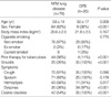
As shown in Table 1, patients with NTM lung disease were of older age (58±14 vs. 50±17 yr, P=0.008), showed a female predilection (82% vs. 26%, P<0.001), and were more likely to have a history of tuberculosis treatment (56% vs. 11%, P<0.001) compared with those with DPB. A past history of or current chronic sinusitis was more common in patients with DPB (100%) than in those with NTM lung disease (26%, P<0.001).
The main presenting symptoms were cough and sputum in both groups. Hemoptysis was more common in patients with NTM lung disease (56% vs. 31%, P=0.017), while exertional dyspnea (97% vs. 26%, P<0.001) and coarse crackles (100% vs. 54%, P<0.001) were more common in patients with DPB.
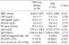
The results of laboratory findings are summarized in Table 2. The WBC counts of peripheral blood and the serum levels of CRP and IgA were significantly higher in patients with DPB than in those with NTM lung disease. The proportion of patients who had a titer of cold hemagglutinin ≥64 was also higher in patients with DPB. The patients with FEV1/ FVC <70%, or arterial oxygenation <80 mmHg were more commonly diagnosed with DPB than NTM.
Comparison of radiographic characteristics
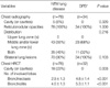
The most common chest radiography finding was the presence of reticulonodular opacities, which were seen in all patients with NTM lung disease and DPB. The laterality and distribution of chest radiography abnormalities were very similar in both diseases; the opacities were bilateral in 90% of patients with NTM lung disease and in all patients with DPB (Table 3).
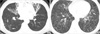
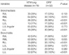
The most common HRCT findings were the presence of bronchiectasis and bronchiolitis, which were seen in all patients with NTM lung disease and DPB. However, the involvement of bronchiectasis and bronchiolitis on chest HRCT was more extensive in patients with DPB. Bronchiectasis and bronchiolitis were usually observed in more lobes in patients with DPB than in those with NTM lung disease (P<0.001, P<0.001, respectively) (Table 3) (Fig. 1). In patients with DPB, the involvement of bronchiectasis and bronchiolitis in the right middle lobe and both lower lobes was also more common than in those with NTM lung disease (Table 4). However, a cavity (or cavities) was more commonly found in patients with NTM lung disease (23% vs. 0%, P=0.003).
DISCUSSION
The purpose of this study was to identify clinicoradiographic differences between the nodular bronchiectatic form of NTM lung disease and DPB. We found that patients with the nodular bronchiectatic form of NTM lung disease were of an older age, had a female predilection, and were more likely to have a history of tuberculous treatment, while patients with DPB had sinusitis more frequently. Hemoptysis was related to NTM lung disease, and exertional dyspnea and coarse crackles were related to DPB. In patients with DPB, marked obstructive abnormalities, hypoxemia, increased inflammatory laboratory findings, and more lobes involved with bronchiectasis or bronchiolitis on HRCT were more frequently observed.
The NTM lung disease has been differentiated into two distinct subtypes, the upper lobe cavitary form and the nodular bronchiectatic form (1, 2). The nodular bronchiectatic form was reported in more than half of NTM lung disease cases and occurs predominantly in non-smoking, middle-aged or elderly women without previous or underlying lung diseases (1, 2). The radiographic findings show bilateral nodular or reticulonodular changes, particularly in the middle lobe of the right lung and the lingular segment of the upper lobe of the left lung (1, 2). In the present study, more than 80% of patients with nodular bronchiectatic form of NTM lung disease had involvement in the middle lobe of the right lung and the lingular segment of the left lung on chest HRCT scans.
Since the first comprehensive report of DPB was published in 1983 (15), there has been dissemination of knowledge about DPB. The diagnosis is based on the clinical and radiographic findings, pulmonary function test, and laboratory findings of patients as shown in the diagnostic criteria of the Ministry of Health and Welfare of Japan (10). Our study evaluated patients who fulfilled these diagnostic criteria. This is the reason nearly 100% show exertional dyspnea, crackles, and sinusitis in DPB. But, even in the nodular bronchiectatic form of NTM lung disease, exertional dyspnea, crackles, and sinusitis were observed in 26%, 54%, and 26% of patients, respectively. As the diagnostic criteria reflect chronic respiratory impairment, other laboratory findings suggest nonspecific inflammation (16). Our study also demonstrated the increased value of WBC, CRP, and IgA in patients with DPB and all of them were significantly higher than those with nodular bronchiectatic form of NTM lung disease. As IgA in immunoglobulin isotype is found predominantly in airway secretion and has a protective effect of the mucosal immune system against respiratory tract infection, increased IgA level in DPB patients might suggest that DPB is more characterized by chronic airway inflammation (17).
A previous study reported that bilateral bronchiectasis, especially combined with multiple small nodules and branching centrilobular nodular structures on chest HRCT, was associated with NTM infections, followed by DPB (7). Our study demonstrated that both groups had similar findings on the chest radiography films, and bronchiectasis and bronchiolitis on chest HRCT without significant differences. However, cavitary lesions were observed only in the nodular bronchiectatic form of NTM lung disease, and the number of involved lobes, especially bilateral lower lobes including the right middle lobe, was greater in DPB patients.
The importance of discriminating between the nodular bronchiectatic form of NTM lung disease and DPB is because of the different treatments for these diseases. In DPB, since the introduction of erythromycin therapy by Kudoh in the mid-1980s, the prognosis of DPB has changed from a fatal to a curable disease (10, 13). Presently, low-dose and long-term erythromycin monotherapy is established as the most efficient treatment, and the clinicians start erythromycin as soon as the patient is diagnosed (10). When erythromycin is found ineffective, the newer macrolides, such as clarithromycin or azithromycin are considered as the second choice.
These macrolides are also important in the treatment of NTM lung disease; especially MAC lung disease and M. abscessus lung disease. In addition to M. kansasii, MAC and M. abscessus are the most commonly encountered pathogens and more than half of patients with MAC or M. abscessus present with the nodular bronchiectatic form on chest CT scans. Introduction of the newer macrolides resulted in a major therapeutic advance in the treatment of MAC lung disease (1, 2). When a macrolide was administered alone, however, the organism was more likely to acquire macrolide-resistance which was strongly associated with a poor treatment outcome (14). Thus, combination therapy including macrolides is recommended for patients with MAC lung disease. In M. abscessus lung disease, macrolides are the only oral antibiotics this organism is susceptible to, and macrolide monotherapy without other intravenous antibiotics, such as cefoxitin, amikacin and imipenem cannot achieve negative sputum conversion (1, 2). Hence, in NTM lung disease, if macrolide monotherapy is prescribed as it is in DPB, it can lead to the acquisition of macrolide-resistance of the NTM infection and even treatment failure. In addition, patients with DPB are recommended for immediate treatment. While, as the course of NTM pulmonary infections of the nodular bronchiectatic form can be indolent, clinicians must determine when to start antibiotic treatment (1, 2).
In conclusion, this study demonstrates that some clinical and radiographic findings are helpful in discriminating between the nodular bronchiectatic form of NTM lung disease and DPB. However, there is considerable overlap in the clinical and radiographic appearances of nodular bronchiectatic form of NTM lung disease and DPB. Still, there is no critical discriminating clinical or radiographic characteristics between them that is as specific as the microbiologic documentation. The correct diagnosis, including aggressive microbiologic evaluation, should be made for the appropriate management of patients presenting with bilateral bronchiectasis and bronchiolitis.
 XML Download