

 PDF
PDF ePub
ePub Citation
 Print
Print


INTRODUCTION
Allergen specific immunotherapy is the only curative treatment modality available at present and has been used successfully in limited allergic diseases, such as insect venom allergy, allergic rhinoconjunctivitis (1, 2) and asthma (3-5). Controlled long-term trials suggest that immunotherapy has the capacity to modify the natural history of allergic airway disease by reducing the incidence of new sensitization (6-8), reducing allergic symptoms through years after discontinuation and preventing the incidence of asthma (1, 2, 4). But the precise molecular mechanisms underlying this treatment modality remain elusive.
Although previous studies were focused on circulating blocking antibodies (9-11), they could not explain the antigen-specific effects of immunotherapy. Other suggested mechanisms were reducing in the numbers of mast cells and eosinophils including the release of mediators (12-14). Recent studies suggest the modulation of interleukin (IL)-4 and/or interferon (IFN)-γ production from allergen-specific T cells (4, 5, 15-19), especially a decrease in allergen-induced T cell activation and both Th2 (IL-4, IL-5, and IL-13) and Th1 cytokines (IFN-γ), which were induced by the increase of regulatory T cell cytokine production, such as IL-10 and/or transforming growth factor-β (TGF-β) (20-23).
The hypothesis of our study was that immunotherapy may induce the functional modification from Th2 to Th1 phenotype in T cells, and these changes will be induced in early phase of rush immunotherapy (RIT) and continued to late phase. To investigate the modifications of cellular immunity in the mechanism of RIT, we evaluated the production of intracellular IL-5 and IFN-γ from the peripheral blood T cells in the early and late phase of the allergic inflammation in asthmatic children with and without RIT.
MATERIALS AND METHODS
Subjects
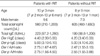
Twenty five children with atopic asthma (16 boys and 9 girls) who were sensitive only to Dermatophagoides farinae (Der f) and Dermatophagoides pteronyssinus (Der p) were enrolled. Patients were divided into two groups, such as those who had received RIT (n=15, RIT group) and those who had never received RIT (n=10, control group).
The diagnosis and severity of asthma were determined for each patient by the American Thoracic Society guidelines (24). Asthma was confirmed by a history of dyspnea and wheezing during the previous 12 months, a greater than 12% reversibility of forced expiratory volume in one second (FEV1) spontaneously or after β2-agonist inhalation, and/or a methacholine provocation test result with a PC20 less than 16 mg/mL. All of the patients were belong to mild to moderate asthma. The two groups were comparable for the severity of asthma, the concentration of total immunoglobulin E (IgE) and Der f or Der p-specific IgE, and allergic skin test to Der f or Der p (Table 1). At the time of the study, all patients were symptom free (for at least 4 weeks) and did not have any medication for asthma, except for short acting β2-agonist, for at least 4 weeks before blood sampling. The ethics committee of the Asan Medical Center Institutional Review Board approved the study, and written informed consents were obtained from the parents of all subjects.
Protocol of rush immunotherapy
The patients received RIT according to the protocol in our pediatric ward. The patients were admitted for 4 days during induction period, and the dosages of each injection were rapidly increased in ten-fold daily starting from 5 Therapeutic unit (TU)/mL. Then they received injections every 2 weeks till 8 weeks until maintenance was achieved (5,000 TU/mL). After then they received injections every 4 weeks. Allergen extracts were obtained from Bencard Allergie (Munchen, Germany).
Measurements of symptom scores and skin reactivity to Der f
The respiratory symptom scores of asthma (25) and the skin test reactivities (allergen/histamine ratio) to Der f and Der p were measured. The severity of symptoms was scored on a Likert scale from 0 to 5 (0=none, 1=trivial, 2=mild, 3=moderate, 4=severe, and 5=very severe). Asthma symptom complaints were divided into three periods: awakening (four items), daytime (six items), and nighttime (five items). The total numbers of nocturnal awakenings for asthma per week were translated into the same six-point scale as follows: no awakening=none, 1 awakening=trivial, 2 to 3 awakenings=mild, 4 to 6 awakenings=moderate, 7 to 10 awakenings=severe, and 11 or more awakenings=very severe.
Lymphocyte proliferation assay
Peripheral blood mononuclear cells (PBMCs) from the patients were centrifugated by Ficoll-Hypaque gradient (Pharmacia, Uppsala, Sweden) and were incubated in 96 well round bottom plate at 106 cells/mL in 200 µL/well. The dosages of Der f allergen (Allergopharma, Reinbek, Germany) were 1, 10, 30, and 50 µg/mL concentration. Proliferative responses were assayed by tritiated thymidine incorporation after 5 day's culture after addition of 1 µCi 16 hr before cell harvesting.
Flow cytometric analysis of PBMCs for intracellular cytokine production
For intracellular T-cell cytokine detection, we employed a flow cytometry, which has been described elsewhere (26). PBMCs (107 cells/mL) were stimulated with phorbol myristate acetate (25 ng/mL; Wako, Osaka, Japan) and ionomycin (1 µg/mL; Sigma Chemical Co., St. Louis, MO, U.S.A.) in the presence of 2 mM/L monensin for 4 hr at 37℃ in a humidified atmosphere of 5% CO2 in air. Cells were fixed in 4% paraformaldehyde and made permeable with saponin. After blocking with 10% AB serum, cells were incubated with a monoclonal mouse anti-cytokine antibody (anti-IL-5 and anti-IFN-γ: Becton-Dickinson, Franklin Lakes, NJ, U.S.A.) at 1 mg/mL in 0.1% saponin. Twenty minutes later, cells were washed twice and incubated with a fluorescein isothiocyanate- or phycoerthrin-labeled polyclonal goat anti-mouse isotype specific antibody and an anti T-cell surface marker antibody (anti-CD3, anti-CD4, and anti-CD8: Becton-Dickinson) for 30 min. Then cells were analyzed by FACSCalibur (Becton-Dickinson) and frequency of cytokine-producing cells in different subpopulation was calculated.
RESULTS
Respiratory symptom scores and skin test reactivity to Der f
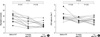
The respiratory symptom scores were decreased significantly 8 weeks after RIT and maintained until 1 yr after RIT (Fig. 1A), but not in control group (data not shown). In addition, the skin test reactivity (allergen/histamine ratio) to Der f was decreased significantly 8 weeks after RIT and maintained until 1 yr after RIT (Fig. 1B), but not in control group (data not shown).
Cellular proliferation assay
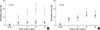
The proliferative response was expressed as stimulation index (SI: cpm with allergen/cpm with medium). The cellular proliferative responses (SI) to each dose of Der f showed dose-dependent responses and the maximum response was found in the stimulation of Der f 30 µg/mL concentration. These responses were decreased significantly 8 weeks after RIT and maintained until 1 yr after RIT (Fig. 2A). However, in the control group the cellular proliferative responses were not suppressed even on 8 weeks and 1 yr follow-up (Fig. 2B).
Changes of T cell population during RIT
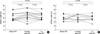
By flow cytometry, the frequencies of CD4(+) and CD8(+) T cells were analyzed before and after RIT. The CD3(+) cells and CD4(+) cells increased at 8 weeks after RIT, but these cells decreased at 1 yr after RIT compare to the cell population at 8 weeks after RIT (Fig. 3A). In contrast, there were no significant changes in 3 follow-up periods in the control group (Fig. 3B). In addition, the CD8(+) cells were not changed at 8 weeks and 1 yr follow-ups in either groups (data not shown).
Intracellular IFN-γ/IL-5 ratio from the peripheral T cells
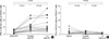
Intracellular IFN-γ and IL-5 staining for CD3, CD4, and CD8 were carried out on activated PBMCs. RIT increased IFN-γ/IL-5 ratio from the CD3(+) T cells (data not shown), and from the CD4(+) T cells at 8 weeks after RIT (P<0.01), but not from the CD8(+) T cells (data not shown), compared to those before RIT. IFN-γ/IL-5 ratio was maintained until 1 yr after RIT from the CD4(+) T cells (P<0.01, Fig. 4A), whereas IFN-γ/IL-5 ratio from the peripheral CD3(+), CD4(+) or CD8(+) T cells was not changed in each follow-up period in the control group (Fig. 4B). These results suggest that RIT induced the modification of cellular immune responses in the peripheral T cells, such as Th2 to Th1 shift in the peripheral CD4(+) T cells.
DISCUSSION
This study shows early increase of IFN-γ/IL-5 ratio from the peripheral blood CD4(+) T cells in children with house dust mite-sensitized asthma even at 8 weeks after RIT. Additionally this change of IFN-γ/IL-5 ratio was maintained till 1 yr after RIT, whereas these findings were not detected in asthmatic children without RIT. These changes were mainly originated from increase of IFN-γ production rather than changes of IL-5 production from the peripheral CD4(+) T cells regardless of RIT. These findings suggest that the immunologic changes after RIT may start early, especially before maintenance therapy, and be originated from the production of the IFN-γ particularly from the CD4(+) T cells, not from the CD8(+) T cells. In addition, these early changes were maintained until 1 yr after RIT. These findings suggest that the Th1 shift on peripheral blood CD4(+) T cells may be one of the main functional modifications of allergen specific immunotherapy and also this effect may be maintained during immunotherapy. Our data is compatible with some of the previous reports (4, 5), whereas some reported that IFN-γ increases while IL-4 diminishes (5, 15, 19) and some others showed that IL-4 was decreased while IFN-γ was not altered (16-18). In addition, others showed a decrease in both cytokines IFN-γ and IL-4 (20, 27). However, these variable results for the immunologic changes after immunotherapy might depend on the protocol of immunotherapy, the type of allergen, the time point of evaluation, the type of stimuli used for cytokine production, and the cell sources used.
The most important change in the allergen-specific immune response during immunotherapy is characterized by a decrease in allergen-induced T cell activation and the production of both Th1 and Th2 cytokines, which outlines peripheral T cell tolerance to allergens. Several studies have shown an increase in regulatory T cells, especially in bee-venom immunotherapy (20, 22, 23). T cell tolerance can be directly initiated by the autocrine action of IL-10 (21, 22), which can inhibit the full maturation of dendritic cell (28). This results in the long lasting induction of tolerance in both Th1 and Th2 cells.
Also, the respiratory symptom were improved significantly in the early phase of RIT only and that was also maintained until 1 yr after RIT. In this study, there was good correlation between the changes of respiratory symptom score and the changes of IFN-γ(+)/CD4(+) cells. In addition, skin reactivity to Der f, which means skin mast cell reactivity, was also significant decrease in the early phase of RIT, and that was persisted until 1 yr after RIT. These findings suggest that not only asthma symptoms but also target tissue responsiveness are improved after RIT, which may be related to the shift of Th1 immune response or decreased histamine release from target cells including basophils and mast cells (14).
IFN-γ-production from peripheral CD4(+) T cells was lower in atopic asthmatics (29). According to these results, immunotherapy may induce the shift of cytokine balance from Th2 to Th1 milieu in the systemic circulation.
RIT suppressed the cellular proliferation in each dose of Der f, and also this suppression was maintained until 1 yr after RIT only in RIT group in contrast to control group. These data were compatible to the previous study (4) which showed decreased proliferation of peripheral blood mononuclear cells to relevant allergen (Der p) in early phase of RIT, but not to irrelevant allergen. Although the inhibition of cellular proliferation underlies the clinical effect of RIT, the mechanism of RIT is not clear. The possibility may be suggested by the induction of allergen-specific suppressor T cells (30), the induction of anergy (31), or the induction of IFN-γ (32).
To confirm the increase of IFN-γ production from the PBMCs after RIT, we also evaluated the serial studies at three different time points during RIT in 7 patients, such as 8 weeks before, immediately before, and 8 weeks after RIT. IFN-γ(+)/CD4(+) T cells were not changed between 8 weeks before and immediately before RIT, but were increased at 8 weeks after RIT (P<0.05, data not shown). These data suggest that IFN-γ(+)/CD4(+) T cells might be increased by the effect of RIT.
The mechanism of increment of IFN-γ producing cells by RIT is not defined. The possible mechanism might be explained by the route of allergen exposure and the amount of administered allergens, which may alter the function of antigen presenting cells (33). The use of repeated high allergen concentrations during RIT may lead the shift to the Th1 cell response. It appears that RIT may correct the decreased IFN-γ production of CD4(+) T cells in the children with atopic asthma.
All our results were originated from the peripheral blood T cells of the asthmatic patients, therefore these did not reveal the direct cause and effect of RIT in the asthmatic airway, so we pointed the systemic effect of RIT. Additionally we suggest that the Th1 shift of immunologic cells may be important in the mechanism of RIT.
 XML Download