

 PDF
PDF ePub
ePub Citation
 Print
Print


INTRODUCTION
Colonic arteriovenous malformation (AVM) is a well-known cause of lower gastrointestinal bleeding (1). Unlike small vascular ectasia or angiodysplasia, they are not restricted to the elderly, tend to be solitary, can be identified endoscopically as flat or elevated bright red lesions (2-4). In addition, they are not confined to the right colon and large in size (3, 4).
Only eight cases of polypoid colorectal AVMs are reported up to date (2-8). Their sizes were between 0.7 cm and 6.2 cm. All patients revealed lower gastrointestinal bleeding. All the lesions were solitary, usually located on transverse, descending and sigmoid colon. They were resected successfully by polypectomy or surgical resection which resulted in the correction of gastrointestinal bleeding (2-8).
Recently, we experienced a case of non-solitary and small cecal AVMs which were found incidentally on endoscopic examination. The lesion was successfully removed by endoscopic biopsy without any complication. To the best of our knowledge, this is the first case of multiple cecal AVM which were found incidentally and successfully removed by biopsy
CASE REPORT
A 66-yr-old woman was referred for routine cancer screening of upper and lower gastrointestinal tract during the admission in the department of Nephrology. She was waiting for arteriovenous shunt operation in the forearm to start hemodialysis. She was suffering from type 2 diabetes, end stage renal disease, hypertension, and anemia of chronic disease. She had neither history of appendectomy nor tuberculosis. Chest radiography on arrival revealed normal finding. She was on insulin, erythropoietin and antihypertensive medications. On admission, her blood pressure was 120/80 mmHg and pulse rate was 80 beats/min. Laboratory examination revealed anemia of chronic disease with hemoglobin level being 11.0 g/dL, Ferrum 177 µg/dL (normal range 65-157 µg/dL), total iron binding capacity 394 µg/dL (normal range 256-426 µg/dL), ferritin 110.4 ng/mL (normal range 13-150 ng/mL), and erythropoietin 7.0 mU/mL (normal range 10.2-25.2 mU/mL).
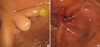
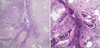
She had no history of gastrointestinal bleeding. Upper gastrointestinal endoscopic finding disclosed no abnormality. Colonoscopy was performed with the aid of Olympus video-colonoscope (CF-H260AI). The entire colonic mucosa appeared normal except for presence of three semi-pedunculated polyps. Two polyps were less than 5 mm in size and were located near to the appendiceal orifice (Fig. 1A). Another 3 mm sized semi-pedunculated polyp was located behind the previous mentioned polyps (Fig. 1B). Since the lesion revealed normal looking epithelium with converging folds, impression of endoscopic finding was inflammatory polyps. Biopsy was taken to confirm the nature, and three biopsies were taken on each lesion. After the biopsies, bleeding from the biopsied site was ceased spontaneously without delay. Histopathologic evaluation of all specimens demonstrated intramucosal hemorrhage and dilated submucosal vessels consistent with polypoid colonic AVM (Fig. 2A). Verhoeff's elastic stain revealed internal laminae within the vessel walls which suggested that these AVMs were consisted of abnormal arteries and veins (Fig. 2B). There was no evidence of gastrointestinal bleeding during the six months of follow-up period after the removal.
DISCUSSION
Histologically, AVMs are believed to be degenerative lesions which are the result of intermittent, low-grade obstruction of submucosal vein as they penetrate the muscular layers of the colon causing small arteriovenous communications (9). Colonic AVMs were also known as vascular ectasia or angiodysplasia (9). Recently, differential diagnosis of these lesions was made by pathological findings (6, 10). Angiodysplasia is mostly found in the cecum and ascending colon, and consisted of mostly venous distensions. Besides, vascular ectasia is consisted of dilated capillary vessels. Dieulafoy's lesion which is also difficult to distinguish from AVMs is an arterial lesion. AVMs have distinguishing histology of ateriovenous vascular abnormality. Clinically, AVMs have distinct clinical manifestations from small vascular abnormalities such as vascular ectasia or angiodysplasia whereas they are not restricted to the elderly, are usually solitary, can be identified endoscopically, are not confined on the right colon, and are large in size (6).
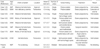
Endoscopically, AVMs generally appear as flat or elevated bright red lesions. A polypoid appearance is extremely rare in the large intestine. There were only 8 cases reported in the English language literature (Table 1). The present case differs from previously reported AVMs in several aspects; 1) small, 2) non-solitary, 3) located on the cecum, 4) covered with normal colored mucosa without erythema, ulcer, or hemorrhagic spots, 5) removed successfully by biopsy, and 4) asymptomatic which were found incidentally (Table 1). To the best of our knowledge, this is the first case of multiple and semipedunculated AVMs in cecum found incidentally by screening colonoscopy.
Due to the rarity of colorectal AVM, most of the cases are diagnosed only after the resection and treatments are not determined yet. Moreover, it is very hard to raise the possibility of a polypoid AVM by endoscopic appearance, that majority are likely to be managed by routine polypectomy (2-5, 7, 8). There was only one case reporting significant bleeding after the removal (6). In the present case, lesions were diagnosed as inflammatory polyps, and were successfully removed by biopsy without significant bleeding.
In summary, we experienced multiple colonic AVMs in cecum which were hard to differentiate from inflammatory polyp, and were successfully removed by biopsy. Our case suggests that small AVM without evidence of bleeding on the surface such as hemorrhagic spot, erythema, or ulcer, could be successfully removed by biopsy without complication.
 XML Download