

 PDF
PDF ePub
ePub Citation
 Print
Print


INTRODUCTION
Pulmonary Arteriovenous Malformations (PAVMs) are unusual abnormalities of the pulmonary vasculature and are often associated with hereditary hemorrhagic telangiectasia (HHT) (1). They lead to a right-to-left shunt due to a direct connection between a pulmonary artery and a pulmonary vein.
Symptoms related to PAVMs often develop between the fourth and sixth decades. Most of the congenital PAVMs (60% to 90%) are associated with HHT (2). Common clinical features are epistaxis, telangiectasias, cyanosis, dyspnea, and gastrointestinal bleeding (3). Neurological complications such as migraine headaches, strokes, transient ischemic attacks, cerebral abscesses, and seizures are most commonly seen (1). The prevalence of migraines in patients with PAVM and HHT seems to be high (4).
Utilizing pulmonary arteriography, arteriovenous malformations can be identified and selectively embolized. However, large-sized single lesions or lesions that are at high risk of complications with transcatheter embolization (TCE) are indicated for surgical resection.
We reported a case of PAVMs in a patient presenting with chronic migraines that was successfully treated with surgical resection.
CASE REPORT
A 41-yr-old woman presented to the emergency room with headache, nausea, dyspnea, and cyanosis suddenly worsening five hours prior to presentation. She reported a history of chronic headaches and dyspnea on exertion of 10 yr duration. She suffered from recurrent left temporo-parietal throbbing headaches that had been poorly controlled by nonsteroidal anti-inflammatory drugs, antidepressants, and minor tranquilizers. Only triptans ameliorated her headaches, but effectiveness lasted only for a few hours. She was aware of neither aura nor any associated symptoms before the headache occurred. She reported no neurologic deficits. The headache occurred 4 to 5 times a month and did not worsen with exertion. Nausea accompanied her headaches and often was followed by emesis with aggravation of the headaches.
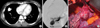
The findings of magnetic resonance image and electroencephalogram were normal. An arterial blood gas study revealed pH 7.49, pCO2 27 mmHg, pO2 69 mmHg, and SaO2 95%. Simple chest radiography revealed a solitary nodule with a relatively definite boundary in the right lung. This was then further characterized as a possible right middle lobe PAVM by computerized tomography (Fig. 1A).
In order to definitively diagnose the lesion as a PAVM and to search for accompanying cerebrovascular malformations, we performed pulmonary arteriography and cerebral angiography. The patient's PAVM was found to be a connection between the artery and vein of the right middle lobe (Fig. 1B). No cerebrovascular malformations were found.
Lung perfusion scan performed prior to surgery revealed perfusions of 48% in the right lung and 52% in the left lung, lending further evidence of a right-to-left shunt in the right lung. Electrocardiogram did not show cardiamorphia or pulmonary hypertension.
The patient's pulmonary arteriogram revealed that the feeding artery of the PAVM had gradually expanded in size. If TCE were to be performed in this case, there would be a high risk of paradoxical embolism caused by unintended migration of a detachable steel coil. Therefore, the decision was made for surgical resection.
A right lateral thoracotomy was performed under general anesthesia. The feeding artery and draining vein measuring about 1 cm in size with a thin pulsatile membrane was found centrally located in the right middle lobe (Fig. 1C). Once the artery and vein were clipped, the PAVM lost its pulsatile beat and decreased in size.
Complete resection of the right middle lobe along with the offending PAVM was performed. Following the operation, the patient's presenting symptoms of migraines, nausea, dyspnea, and cyanosis were resolved and arterial blood gas study improved to pH 7.47, pCO2 33 mmHg, pO2 86 mmHg, and SaO2 97%. The patient was subsequently discharged and now lives a healthy life without evidence of recurrence over the past 10 months.
DISCUSSION
Although approximately 70% of the cases of PAVMs are associated with HTT, our patient had no personal evidence or family history of HHT and was diagnosed with isolated congenital or sporadic PAVM.
The frequency of migraine headaches in patients with sporadic PAVMs is unknown. The presentation of our patient with migraines that resolved after surgical resection of her PAVM suggests that the etiology of her migraines was related to the PAVM. Increased prevalence of migraines has been described in patients with HHT, the most common cause of PAVMs (5). Approximately 30% of people with HHT have PAVMs, leading to pulmonary right-to-left shunt (6). Two studies recently showed an increased prevalence of migraines in patients with pulmonary right-to-left shunts (PAVMs) (7, 8).
A causal relationship between the presence of a right-to-left shunt and migraines has been suggested but remains unproven until now. Several hypotheses have been suggested. Firstly, HHT and some subtypes of migraines are autosomal-dominant disorders. It is possible that a particular genetic substrate that may determine pulmonary right-to-left shunt in patients with HHT and may also activate migraines (9). Secondly, trigger substances might enter the systemic circulation through the right-to-left shunt instead of being trapped in the pulmonary capillaries. These trigger substances might induce cerebral vascular instability or increased excitability of central nervous system and cause migraine headaches. Trigger substances that are implicated are vasoactive chemicals such as serotonin or microemboli (10).
Treatment until 1980 was limited to surgical ligation and resection, but improved catheterization techniques have made embolotherapy the treatment of choice for most patients (11).
Utilizing pulmonary arteriography in patients with PAVMs, the origin of the artery that enters into the lesions can be identified and subsequent treatment method can be tailored to the patient's clinical condition and the characteristics of the PAVMs including size, number, location, and treatment complications. Historically, indications for surgical intervention for PAVMs include gradual expansion of the lesion and paradoxical embolism (12). As in our case, solitary PAVMs 2 cm or larger and centrally located are also indicated for surgery.
Two studies recently showed a high prevalence of migraines, 43 to 59%, in patients who were admitted for TCE of large PAVMs (4, 5). These two groups, however, did not study the effects of embolization on the prevalence of migraines. Only one study has reported a significant reduction in the prevalence of migraines after TCE of PAVMs (13).
However, there has not been a study that describes significant reduction in the prevalence of migraines after surgical operation of PAVMs. We report a case of a patient whose migraines resolved following successful surgical resection of a sporadic PAVM. Further studies are needed on a population basis to determine whether surgical resection of PAVMs reduces the prevalence of migraine headaches in this unique population.
 XML Download