

 PDF
PDF ePub
ePub Citation
 Print
Print


INTRODUCTION
There are many treatment options for cervical disc disease. The standard treatment of choice is anterior cervical discectomy and fusion (ACDF). Although most patients wish to treat both the posterior neck pain and associated arm pain, some patients wish to be treated with less invasive methods, even though some tolerable symptoms may persist, due to their social/physical situations. Here we present one method of treatment for socially/physically active patients.
MATERIALS AND METHODS
Patients
Three consecutive patients underwent endoscopic posterior cervical discectomy between November 2007 and Januaray 2008. All of the patients primarily complained of radiating arm pain, which was associated with a tolerable degree of neck pain. All of the patients were middle-aged soldiers with families. In Korea, only physically healthy persons are allowed to serve in the military, and if a soldier was to undergo cervical spine surgery with implantation, the soldier discharged from the military against their will according to the military regulation. It is very difficult for middle-aged soldiers to get new jobs. A pension after retirement is not enough to provide for a middle-aged father and his family. If they want to keep their job after undergoing surgery, they should be able to perform their military drills after the operation.
Therefore, many doctors have agonized over how to treat middle-aged soldiers without forcing them to sacrifice their jobs and how to help them to maintain a level of physical fitness that is sufficient for soldiers. Posterior cervical endoscopic discectomy was performed in all three patients in the present study.
The first patient was a 42-yr-old sergeant. He had been suffering from radiating pain in the left C7 sensory dermatome (SD) for 2 yr. The pain intensity, measured by the visual analogue scale (VAS), was 8/10. He had been waiting for a promotion and endured the pain for 2 yr in hopes of getting the promotion. He is the father of a high school student. The severity of the associated neck pain was VAS 3/10. Physical examination revealed weakness in the right triceps (motor grade IV/V), and triceps jerk could not be elicited. Magnetic resonance imaging (MRI) revealed disc protrusion in the right C6-7 foramen associated with disc degeneration at C5-6 (Fig. 1A).
The second patient was a 46-yr-old sergeant major. Although he had already been promoted, he wanted to keep his job until he reached the retirement age of 54 yr. He has one son and one daughter, both of whom are in high school. He is a healthy man, and he did not want to be discharged from the army while in his socially and physically active stage of life. He complained of radiating pain at the left C7 SD (VAS 7/10) and posterior neck pain (VAS 2/10) that had lasted for 2 months. Physical examination revealed weakness in the left triceps (motor grade IV/V), and triceps jerk could not be elicited. MRI showed foraminal disc protrusion on the left at the C6-7 level.
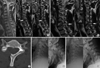
The third patient was a 50-yr-old sergeant major. He had been suffering from severe radiating arm pain at the left C7 SD (VAS 10/10) for 2 weeks. The severity of the associated neck pain was VAS 4/10. Physical examination revealed weakness in the left triceps (motor grade III/V), and triceps jerk could not be elicited. MRI showed disc rupture on the left posterior lateral side of the C6-7 disc space (Fig. 2A). He is the father of a son and a daughter. He had devoted his whole life to the army. He wanted to treat the arm pain first, but he also wanted to keep his job as a soldier. He really wanted to retire with glory and not disease.
All operations were performed after listening to the patients' needs and explaining the anticipated medical and social/physical outcomes. The patients were followed-up for more than 2 months after the operations. MRI was performed one week after the operations. During the follow-up period, motor grade was evaluated and flexion/extension films of the cervical spine were also checked.
Operations
All operations were performed under general anesthesia. Each patient was situated in the prone position, and the neck was flexed without fixation. The operation was similar to traditional foraminotomy (1, 2). The entry point was 1 cm from the midline and just above the post-articulation. The incision length was approximately 1 cm. Gentle palpation of the inter-laminar space was made possible after the obturator (6.9 mm outer diameter) was introduced. The working channel was introduced into the obturator, and the obturator was retrieved. The opened bevel of the working channel was directed toward the medial side in order to avoid accidentally compressing the spinal cord. Drilling was performed from the medial to lateral margin of the inter-laminar space, and the superior facet was drilled first. The ligamentum flavum was removed, and the lateral margin of the dura and exiting root was visualized. Bone drilling could be extended as desired according to the pathology. Fragmentectomy was the only procedure to be performed without internal decompression of the disc. Decompression was confirmed by the lack of compressed lesion inferior and superior to the nerve root. The skin was closed with a single stitch. All of the instruments used in the operations and the video system were manufactured by WOLF (Richard Wolf GmbH, Knittlingen, Germany).
RESULTS
The ruptured fragments were successfully removed in all three patients. Trimming of the protruded annulus was also performed after nucleus fragment removal in the first patient. After the operation, a computed tomography scan was obtained in order to determine the extent of the foraminotomy and to evaluate facet joint violation. In all patients, facetectomy was limited to less than 50% of the medial side (Fig. 1C). The patients were required to wear a neck brace for 7 days, and free neck motion was allowed from the 8th day after operation after the stitches were removed. The first and second patients showed significant improvement in radiculopathy within 1 day after the surgery, and both patients reported more than 90% improvement from the preoperative state, with slight improvements in neck discomfort (excellent outcome by Macnab's criteria). The weakness in their triceps improved to motor grade V/V within 7 days after the operation. Follow-up MRI showed that the protruded disc was successfully removed (Fig. 1B). Dynamic film of the cervical spine taken one week after the operation showed no instability or change in disc height with preservation of motion (Fig. 1D, E). They returned to their military base and were able to perform their military drills without difficulty within 10 days and 1 month of the surgery, respectively.
The third patient complained of residual radicular pain (VAS 4) 1 day after the operation. During the operation, a large disc fragment (1×0.8 cm) was removed, and root decompression was verified. The direction of the herniated nucleus was posterior and lateral, and it was impossible to trim the annulus (capsule of ruptured nucleus) on the medial side because the spinal cord was not retractable. On follow-up MRI, although the ruptured disc was removed, the thick residual of the annulus that formed a capsule around the herniated nucleus remained, and the nerve root was still compressed (Fig. 2B). He also complained of myofascial pain around the left scapula. The patient was comfortable since the severe arm pain in his arm had disappeared, and further rehabilitation was continued one week after the operation. Dynamic film of the cervical spine taken one week after the operation showed no instability or change in disc height with preservation of motion. Trigger point injection relieved most of the myofascial pain around the shoulder and some of the arm pain. The weakness in his left triceps improved (motor grade V/V) within two weeks of the operation. His symptoms improved over time, and the severity of his arm pain was VAS 1 (excellent outcome by Macnab's criteria) 2 months after the operation. Follow-up MRI performed two months after the operation showed slight shrinkage of the capsule (Fig. 2C). He returned to his base two months after the surgery, and he is currently able to perform his military duties without difficulty.
DISCUSSION
There are several treatment options for patients with cervical disc herniation when the pathology is located on the lateral or posterior lateral side and the primary symptom is radiculopathy. The first treatment option is traditional ACDF, followed by cervical arthroplasty, anterior cervical endoscopic discectomy, anterior transcorporeal or transuncal anterior cervical discectomy, traditional posterior microforaminotomy with or without tubular retractor, and posterior endoscopic discectomy. All of these procedures have their own indications and anticipated results.
ACDF is a standard technique with an established result. However, this mode of treatment requires an anterior approach and fusion of the diseased segment, which leads to motion limitation and increased adjacent segment stress/degeneration (2-7). Some authors have reported that the rate of complications, such as recurrent laryngeal nerve injury or dysphasia, was higher than expected (8, 9). Implantation could be burdensome to physically and socially active people, such as soldiers because of the military regulation. Despite the development of improved operative procedures, postoperative scars on the anterior neck also could be a cosmetic handicap for socially active individuals.
The artificial disc was invented to preserve the motion of the diseased segment and to reduce the amount of stress on the adjacent segment (3). However, biomechanical studies have shown that artificial discs are not a true substitute for original discs, as the range of motion on the implanted segment is greater than normal physiologic motion and the motion of the adjacent segment is mostly decreased (3). Moreover, the cost of this procedure is very high in comparison to other procedures. Until now, the artificial disc has not been a true solution for the replacement of human discs. As with ACDF, an implantation could be burdensome to physically and socially active people. The risk of approach-related complications and the cosmetic results could be similar to those of ACDF. There are several other options for removing the pathology while preserving functional motion of the involved level.
Anterior cervical endoscopic discectomy is a good choice for the treatment of lateral disc protrusion. This operation can be performed under local anesthesia and can be used to preserve the motion of the involved level (5-7). Excellent results were achieved in more than 90% of patients (5-7). Ahn and Lee et al. reported that further instability did not occur, even though the height of the disc was decreased (5-7). However, this procedure requires access through the disc anterior to the pathology with violation of the anterior structure, such as the annulus and nucleus. This procedure may eventually reduce the stability of the disc. Radiological confirmation is necessary during the approach, and the operation cannot be performed if the C-arm does not show an involved level, such as the C6-7 or C7-T1 level (5-7). There is also a risk of major vessel complication or nerve injury with this approach (5-7, 10).
Anterior transuncal microforaminotomy, developed by Jho has a patient satisfaction rate higher than 95%, with functional preservation of the involved level (11, 12). However, the problems of disc space narrowing and anterior column violation still remain, even though there were no symptoms (2, 4-7). The transcorporeal approach was developed to solve the problem of uncovertebral joint violation (4, 13). Comparative study of the anterior transcorporeal approach and transuncal approach revealed that patients subjected to the anterior transuncal approach showed a significant decrease in disc height during the follow-up period in comparison to those subjected to the transcorporeal approach (13). Although different operations performed by different surgeons may have different results, the removal of an uncovertebral joint or the medial wall of the foramen exposing the vertebral artery might be a problem. Although the transcorporeal approach could resolve this problem, this procedure also has the risk of complication during the anterior approach and leaves a scar on the anterior side of the neck (4, 13).
Posterior endoscopic discectomy was first introduced in 1999. The procedure is similar to traditional foraminotomy, except that an endoscope is used instead of a microscope, with or without a tubular retractor (1, 2). Rutten et al. published their 2-yr results and reported a success rate of approximately 96% (2). A laterally localized herniated cervical soft disc without instability is the primary indication (2, 14, 15). The complication rate was very low, and there was no operationinduced neck pain or instability (2). Contrary to the previous suggestion (4, 16), direct removal of the offending pathology is possible without violation of the nucleus or bone of the anterior column (2, 4, 15). Moreover, this procedure is possible, even at the C7-T1 level, with direct visualization during the approach (2, 15). The greatest difference between this procedure and conventional posterior foraminotomy is the lack of access-induced muscle injury (1, 2, 14-16). Muscle injury can also be reduced by microforaminotomy with the use of a tubular retractor system (1, 14, 16). There is hardly any difference between the operative skills necessary for this procedure and endoscopic discectomy, with the benefit of less traumatization of the paraspinal muscle (1, 2, 14, 16). The difference between the cosmetic result and accessinduced muscle pain associated with those two procedures may be regarded as minimal. However, the most distinguishing feature of endoscopy is its excellent magnification and illumination (2). Minimal manipulation of the root, facet joint and spinal cord are possible with the help of this feature of endoscopy (2, 15). However, although minimal, there is also the risk of disc narrowing, which occurs in 32% of patients without symptoms or instability (2). This procedure is not optimal for the removal of a centrally located pathology, hard disc or spur (2, 4, 15).
A single method of operation would not be sufficient for all types of cervical disc pathology. There are merits and demerits associated with each procedure. A recent trend in disc surgery is 'functional preservation' with a minimally invasive technique, and posterior endoscopic discectomy may be a good alternative choice for this reason (1, 2, 4-7, 11, 12, 14). The authors expect that stability will be maintained in the present cases because there was no violation of components in the anterior column and the extent of facetectomy was minimal (1, 15, 16). Further comparative study with a longer follow-up period in a larger patient group is warranted.
In conclusion, the gold standard technique for the treatment of cervical nucleus herniation has been anterior cervical discectomy and fusion, until now. Even though the follow-up period in the present study was short, considering the physical/social activity levels and anticipated physical/social loads in some patients such as soldiers, posterior cervical endoscopic discectomy may be a promising alternative for selected cases.

 XML Download