

 PDF
PDF ePub
ePub Citation
 Print
Print


The use of highly active antiretroviral therapy (HAART) in the treatment of human immunodeficiency virus type 1 (HIV-1)-infected individuals has dramatically changed the clinical outcome of many infected persons, contributing to the current substantial declines in both the incidence of AIDS and AIDS-related mortality (1). However, replication-competent virus, HIV-1 proviral DNA, spliced and unspliced HIV-1 RNA in CD4+ T cells, and unidentified viral reservoirs have all been observed in most infected individuals, even when plasma viremia has been suppressed below detectable levels (2, 3). In addition, HAART does not prevent replication in cells harboring competent HIV-1 proviral DNA. These facts underline the impossibility of eradicating HIV-1 using HAART alone (2, 4).
The quantitative determination of HIV-1 proviral DNA load offers significant therapeutic information, especially when HIV-RNA levels drop below detectable limits during HAART treatment (5). However, the clinical factors associated with proviral DNA load have not been elucidated. Some factors, such as CD4+ T cell counts or the CD4+:CD8+ T cell ratio, represent potential clinical variables associated with HIV-1 proviral DNA load (6).
The objective of this study was to evaluate clinical factors associated with HIV-1 proviral DNA load in HIV-1-infected individuals on HAART who have undetectable plasma viral RNA.
Thirty-six HIV-1-infected persons who had been admitted to a tertiary-care teaching hospital were enrolled in this study after they gave informed consent. All subjects were chronically HIV-1 infected patients with undetectable plasma viral loads on HAART. We reviewed the medical record of each patient retrospectively, collecting age, sex, CD4+ T lymphocyte counts, CD8+ T lymphocyte counts, levels of plasma viral RNA, regimens and durations of antiretroviral treatments, and opportunistic diseases, among others. Data at various times of each patients were collected. The collected data contained the mean level of plasma viral RNA when CD4+ T cell counts were above 500 cells/µL without HAART. HIV RNA levels were measured by nucleic acid sequence-based amplification using a NucliSens®fiEasyQ Analyzer (Biomerieux, Boxtel, Netherlands). CD4+ and CD8+ T lymphocyte counts were calculated by multiplying the number of lymphocytes measured with an automatic cell counter by the percentage of CD4+ or CD8+ antigen-positive cells quantified with monoclonal antibodies (Becton-Dickinson, New Jersey, U.S.A.).
Peripheral blood mononuclear cells (PBMCs) were obtained by Ficoll-Hypaque density gradient centrifugation. CD4+ T cells were isolated from PBMCs using a human CD4 cell-separation kit (EasySepTM, StemCell Technologies, Vancouver, Canada). Real-time polymerase chain reaction (PCR) was used to determine the number of HIV-1 proviral DNA copies per 106 PBMCs, as described elsewhere (6). The primers 5'-GGTCTCTCTGGTTAGACCAGAT-3'(5' primer) and 5'-CTGCTAGAGATTTTCCACACTG-3'(3' primer) were used, along with the fluorescent probe, 5'-6FAM-AGTAGTGTGTGCCCGTCTGTTTAMRA-3'. PCR conditions consisted of a denaturation step at 95℃ for 3 min, followed by 45 cycles of 15 sec at 95℃ and 1 min at 58℃. Serially diluted ACH-2 DNA was also subjected to PCR, as above, to obtain standard curves.
The independent t-test and Spearman's rank correlation were used to measure the correlation between HIV-1 proviral DNA load and immunologic, virologic, and clinical parameters. To identify independent relationships between HIV-1 proviral DNA load and various factors, multivariate linear regression analysis was performed with age, gender, CD4+ T cell count, and the variables that had a significant correlation with HIV-1 proviral DNA load on univariate analysis. All p values were 2-tailed, and p<0.05 was considered statistically significant. All analyses were performed using SPSS for Windows 12.0 (SPSS, Chicago, IL, U.S.A.).
The mean age of enrolled patients was 42.5±9.3 yr. Among the patients, 47.2% were AIDS patients. The mean follow-up duration of the subjects was 1,330±1,178 days, and the years of diagnosis of HIV infection were 1991, 1994, 1997, 1999, 2000, 2001, 2002, and 2003 in 1, 2, 2, 5, 3, 2, 6, 8, and 7 patients, respectively. The mean CD4+ T cell count was 431±201 cells/µL, and the mean value of the mean plasma viral RNA level when the CD4+ T cell count was above 500 cells/µL without HAART was 103,168±179,952 copies/mL.
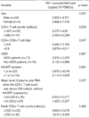
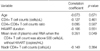
The mean HIV-1 proviral DNA load in all subjects was 2,968±4,956 copies/106 PBMCs. Among the examined clinical parameters, only the mean level of plasma viral RNA when the CD4+ T cell count was above 500 cells/µL without HAART was significantly associated with proviral DNA load (Table 1, 2, p<0.05). There was no significant correlation between proviral DNA load and CD4+ T cell count or duration of HAART. Other clinical factors such as CD4+: CD8+ T cell ratio, nadir CD4+ T cell count, age, or sex were also not associated with HIV-1 proviral DNA load. Multivariate linear regression analysis revealed the mean level of plasma viral RNA when the CD4+ T cell count was above 500 cells/µL without HAART was significantly associated with proviral DNA load (β=0.440, p=0.014).
The qualitative and quantitative evaluation of both serum HIV-1 RNA genome and HIV-1 proviral DNA are pivotal markers in the diagnosis and prognosis of HIV-1 infection (7, 8). Although the quantitative determination of plasma HIV RNA copies directly represents viral replication and is the main prognostic parameter for disease progression (9), the HIV-1 proviral DNA load represents the infection reservoir in PBMC and lymphoid tissues and plays a pivotal role in immune surveillance escape (2, 10). The amount of proviral DNA might also be an important virological marker for exploring viral reservoirs and assessing the impact of treatment (5). Moreover, the presence of this reservoir indicates the possibility of a viral replication rebound when therapy is interrupted or discontinued (2, 10).
In one study, a significant inverse correlation was demonstrated between the frequency of HIV-1 proviral DNA-bearing CD4+ T cells and CD4+ T cell count (11). A similar pattern was discovered for the CD4+:CD8+ T cell ratio of HIV-1 infected individuals receiving HAART in whom plasma viremia had been suppressed below the limit of detection for prolonged periods of time (6). CD8+ T cells also appear to exhibit potent suppressive activity against HIV replication in the latent viral reservoir via direct cellular contact in patients who are naturally long-term nonprogressors or in those treated with HAART (12). Other antiviral activities of CD8+ T cells may be responsible for the suppression of HIV replication in the resting CD4+ T cell reservoirs (12).
In this study, there was a significant correlation between proviral DNA load at the time of undetectable plasma HIV RNA with HAART and the mean level of plasma viral RNA when the CD4+ T cell count was above 500 cells/µL without HAART. The mean levels of plasma viral RNA along with CD4+ T lymphocyte counts above 500 cells/µL could reflect the size and quantity of viral reservoirs.
The threshold level of plasma viral RNA for the prediction of progression of HIV infection is not well defined. We arbitrarily chose 50,000 copies/mL as the cut-off, because in this study a plasma viral RNA load of 50,000 copies/mL effectively divided the subjects into two groups with higher or lower plasma viral RNA.
This study is limited by the fact that it features cross-sectional findings in a small number of subjects. We collected retrospective data without knowing the date of seroconversion, and so we could not consider certain factors that affect the progression of HIV infected patients for multivariate analysis. And, the mean level of plasma viral RNA when the CD4+ T cell count was above 500 cells/µL could be influenced by the progression patterns of subjects and the frequency of the test, and it could not reflect the progression patterns of each patient.
In conclusion, we found evidence that the mean level of plasma viral RNA when the CD4+ T cell count was above 500 cells/µL without HAART may be associated with HIV-1 proviral DNA load at the time of undetectable plasma HIV RNA with HAART, potentially representing the size of viral reservoirs. Strategies to reduce the levels of plasma viral RNA when CD4+ T cell counts are above 500 cells/µL without HAART could help reduce HIV-1 proviral DNA loads.
 XML Download