

 PDF
PDF ePub
ePub Citation
 Print
Print


INTRODUCTION
The metabolic syndrome (MetS) is a clustering of metabolic risk factors, including abdominal obesity, high blood pressure (BP), high triglyceride (TG) levels, low levels of highdensity lipoprotein (HDL) cholesterol, and high levels of fasting plasma glucose (1). The MetS is associated with subsequent increases in the incidence of type 2 diabetes mellitus (DM) (2), cardiovascular disease (CVD) morbidity (3) and even mortality (4).
The MetS is significantly associated with increased levels of alanine aminotransferase (ALT), a liver-specific enzyme (5). Nonalcoholic fatty liver disease (NAFLD) is the hepatic manifestation of the MetS (6). The close relationship between the MetS and NAFLD might be attributable to an overaccumulation of stored fat in the abdomen and liver (7). A previous study showed that the prevalence of the MetS was influenced by an increase in physical activity (8). However, the previous studies only focused on the MetS (8) and were performed only in men (7) or in obese elderly subjects (9).
In the present study, we sought to determine the prevalence of NAFLD and the MetS, and to evaluate the distribution of NAFLD (and its grades of hepatic steatosis) according to the addition of the components of the MetS in relatively healthy hospital workers. We then investigated how visceral fat content and physical activity were associated with both NAFLD and the MetS in men and women.
MATERIALS AND METHODS
Study participants
Potential participants for this study were recruited in 2005 from a group of hospital workers aged 30 to 59 yr at Chonbuk National University Hospital, Jeonju, Jeonbuk, Korea. Age- and sex-stratified random cluster sampling was adopted for the selection of the study population. The hospital workers were comprised of 249 men and 529 women, and a total of 334 (43%) workers were randomly selected for participation in this study. All participants gave written informed consent, and the study was approved by the Chonbuk National University Hospital research ethics committee.
Data collection and measurement
The study was conducted by medical personnel from the hospital, all of whom were trained and supervised by physicians. A blood sample was obtained from each participant. All participants were interviewed using a questionnaire designed to determine whether there was a prior history of CVD, type 2 DM, hypertension, and medication usage. The questionnaire also included questions related to lifestyle, including alcohol-drinking status, as estimated by the frequency, duration, amount and type of liquor consumed. The mean ethanol intake per day was calculated from this data. The participants were divided into three groups according to smoking status: current smokers, ex-smokers, and nonsmokers. Physical activity was investigated using Baecke's Habitual Activity Questionnaire (10), which was translated into a Korean version and validated in a previous study (11). The Baecke's questionnaire was divided into three indexes: 1) physical activity at work, work index; 2) athletic activity during leisure time, sports index; and 3) physical activity during leisure time excluding athletic activity, leisure time index.
Blood pressure was measured on the right arm with the subject in a sitting position using a standard mercury manometer after at least 5 min of rest. Two readings were obtained from each participant at 5 min intervals, and the mean value of the two readings was taken as the individual's BP. Waist circumference (WC) was measured midway between the lower rib margin and iliac crest. Skinfold thickness was measured at the biceps, triceps, subscapular, and suprailiac skinfolds using a Skyndex caliper (Skyndex I Electronic Fat Calipers, Caldwell, Justiss & Co., Inc., AR, U.S.A.). All measurements were performed twice, and the observers were supervised by a single trainer. The Durnin-Womersley formula was used to calculate the percentage of body fat (12), which was closely related to the amount of subcutaneous adipose tissue (13).
Blood samples were collected in the morning after an overnight fast. Serum samples were obtained after venous blood collection and centrifugation at 3,000 g for 15 min. Serum biochemistry analyses were performed on a Hitachi 7600-110 analyzer (Hitachi High-Technologies Corporation, Tokyo, Japan). Both serum aspartate aminotransferase (AST) and ALT activities were measured according to the International Federation of Clinical Chemistry (IFCC) reference method (ASAN Pharmaceutical Co., Ltd, Seoul, Korea). γ-Glutamyl transferase (GGT) was measured using a modified IFCC method (Wako Pure Chemical Industries, Ltd., Osaka, Japan). Serum TG, glucose and uric acid concentrations were determined enzymatically (Roche Diagnostics GmbH, Mannheim, Germany). HDL-cholesterol was measured enzymatically as cholesterol after selective disruption of HDL only (Daiichi Pure Chemicals Co., Ltd., Tokyo, Japan). C-reactive protein (CRP) was determined by a latex agglutination immunoassay using antihuman CRP mouse monoclonal antibody-coated latex (Daiichi Pure Chemicals Co., Ltd.). The level of apolipoprotein B (apoB) was determined by a Roche/Hitachi Modular P Chemistry analyzer using an immunoturbidimetric assay (Roche Diagnostics GmbH). Serological markers of hepatitis B (HBsAg, anti-HBs) were determined by electrochemiluminescence immunoassay (MODULAR ANALYTICS E170, Roche Diagnostics GmbH), and anti-HCV status was determined using a chemiluminescent microparticle immunoassay (ARCHITECT, Abbot Laboratories, Abbot Park, IL, U.S.A.). Daily quality control of the above measurements made on Hitachi instruments was carried out at each site by duplicate measurements with a commercially available control material (Bio-Rad Laboratories, CA, U.S.A.). Assay performance was monitored regularly by the Korean Association of Quality Assurance for Clinical Laboratory and the Korean Society of Laboratory Medicine (KSLM) Laboratory Inspection and Accreditation Program (IAP) (14).
All ultrasonographic examinations were performed by one radiologist (Y.K.K) who had 9 yr of experience performing abdominal ultrasonography. The total number of sonograms performed by Y.K.K. has exceeded 5,000 per annum over the past 8 yr. The radiologist was blinded to the patients' medical histories and their laboratory findings. For each subject, the severity of fatty liver disease was evaluated, and visceral fat thickness (VFT) was measured using a 3.5-MHz convex probe (Sequoia, Siemens Medical Solutions, Mountain View, CA, U.S.A.). The severity of fatty liver disease was subjectively rated by the radiologist based on the following three grading scales (15, 16): grade 1, a slight diffuse increase in fine echoes in the hepatic parenchyma with normal visualization of the diaphragm and intrahepatic vessel borders; grade 2, a moderate diffuse increase in fine echoes with slightly impaired visualization of the intrahepatic vessels and diaphragm; and grade 3, a marked increase in fine echoes with poor or no visualization of the intrahepatic vessel borders, diaphragm, and posterior portion of the right lobe of the liver. In addition to the severity of fatty liver disease, the examiner also assessed the evidence of chronic hepatitis or liver cirrhosis, including hepatic nodularity, coarseness of liver parenchyma, and splenomegaly. VFT was measured along the length of an imaginary vertical line between the posterior aspect of the anterior abdominal wall and anterior line of the abdominal aorta, at the mid-point between the transverse portion of the duodenum and the iliac bifurcation of the aorta. The level that was devoid of bowel or its contents was chosen for the measurement of VFT in order to obtain the purest possible measurement. To avoid any calculation bias, mean values of each of three measurements were acquired. Repeated measurements on the same subjects (12 men and 13 women) gave coefficients of variation (CV) of <1% for the severity of fatty liver disease and <6.5% for VFT.
The MetS was identified by the presence of three or more components of the MetS according to the Third Adults Treatment Panel (ATP-III) of the National Cholesterol Education Program (NCEP) (1), with modified WC cutoff values for Asian populations (17): 1) abdominal obesity (WC ≥90 cm for men and ≥80 cm for women); 2) high TG (≥150 mg/dL); 3) low HDL-cholesterol (<40 mg/dL for men and <50 mg/dL for women); 4) high BP (≥130/85 mmHg or subjects using antihypertensives); and 5) high fasting glucose (≥110 mg/dL).
The exclusion criteria for NAFLD used in the present study were guided by a previous report (18): 1) an alcohol intake of more than 20 g/day, 2) seropositivity for hepatitis B surface antigen (HBsAg), 3) seropositivity for anti-hepatitis C virus antibody (anti-HCV), 4) a prior history of taking medication that could cause steatosis, including steroids, amiodarone, estrogens, etc, and 5) a prior history of any kind of liver disease. Finally, using the current exclusion criteria, we chose the subjects with NAFLD from among the participants with hepatic steatosis.
Statistical analysis
For all variables, the mean (±SD) or proportion was reported according to the number of MetS components. The variables were compared by analysis of variance (ANOVA) or χ2 test for trend, as appropriate. A χ2 test was performed to assess trends in the association between the grade of NAFLD and the number of MetS components. The mean values of VFT according to the grades of NAFLD were presented for each gender, and interaction terms was calculated by two-way ANOVA. According to the presence of NAFLD, independent t-test or χ2 test was performed in the subjects with more than two components of the MetS because NAFLD was found markedly increased from the subjects with just two components. All independent variables that showed differences of more than borderline significance (p<0.1) for NAFLD were entered in a multivariate logistic regression model in order to determine the independent association with NAFLD. All statistical calculations were performed using Stata 8.2 software (Stata Corporation, College Station, TX, U.S.A.).
RESULTS
Among the eligible subjects (n=334), 135 men and 158 women completed the survey (response 87.7%). A total of 69 participants were excluded: 47 (14.1%) men reported consuming more than 20 g of alcohol per day, 11 (3.3%) were seropositive for hepatitis B antigen, and 5 (1.5%) were seropositive for anti-HCV antibody, one (0.3%) was a moderate alcohol drinker and HBsAg positive, and one (0.3%) was anti-HCV positive and an alcohol drinker. In addition, four participants (1.2%) were not subjected to an abdominal ultrasonographic examination, and two of them reported having a history of drug-induced liver disease. A total of 78 men and 146 women (67.1%) were included in the final analysis. The participants and non-participants did not differ significantly with respect to age (mean±SD, 43.8±7.2 and 43.6±7.3 yr, respectively). However, the participants included more women than the non-participants (65.2 and 17.4%, p<0.001).
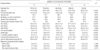
The MetS was observed in 26 subjects (11.6% in total, 20.5% and 6.8% in men and women, respectively, p=0.002), as shown in Table 1. There were 70 subjects with no component of the MetS, 73 with one, 55 with two, 18 with three, and 8 with four or more MetS components (31.3, 32.6, 24.6, 8.0, and 3.6%, respectively). VFT increased significantly with the addition of components of the MetS (p<0.001). In addition, age, past medical history, AST, ALT, GGT, apoB, uric acid, and Durnin-Womersley percent body fat were also significantly increased with the addition of MetS components. The proportion of women and years of formal education decreased significantly with the addition of MetS components. There were no significant changes in smoking, alcohol non-drinking, CRP, and Baecke's physical activity indexes.
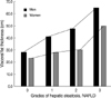
VFT increased significantly according to the severity of NAFLD in both sexes (p<0.001, Fig. 1). Even though VFT was much greater in men than in women for all grades of hepatic steatosis, the interaction terms between sex and grade of NAFLD was not significant (p=0.303, R2=0.627).
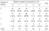
The severity and incidence of NAFLD was significantly associated with the addition of MetS components, as shown in Table 2 (p<0.001). NAFLD was observed in 41.5% of the subjects (57.7% and 32.9% in men and women, respectively, p<0.001). Many subjects with the MetS had NAFLD (73.1%), and components of the MetS were present in 20.4% of the subjects with NAFLD (p=0.001). Thirty-six of the subjects with two MetS components (65.5%) had NAFLD, and 19 (73.1%) of the subjects with more than three components of the MetS also had NAFLD. None of the patients with more than 4 components of the MetS had a normal liver. None of the patients showed any evidence of chronic hepatitis or liver cirrhosis, such as hepatic nodularity, on ultrasonography.
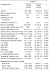
In the subjects with more than two components of the MetS, WC, TG, ALT, AST, GGT, and VFT, were significantly higher in the group of patients with NAFLD than the group without NAFLD (Table 3). In addition, the patients with NAFLD had significantly lower work index scores. Female gender, alcohol non-drinking, apoB, and uric acid showed borderline significance (p<0.1). After adjustment for all potential confounders, NAFLD was independently and positively associated with VFT (OR, 1.10 [95% CI, 1.01-1.19]) and inversely associated with work index (0.29 [0.10-0.87]) (Table 4).
DISCUSSION
This study shows that increased visceral fat content is significantly associated with both NAFLD and the MetS and that there is a significant linear trend in the grade of hepatic steatosis and components of the MetS. Increased VFT can be a common denominator of both NAFLD and the MetS because VFT showed an independent association with NAFLD, even in the subjects with more than two components of the MetS. In addition, the negative association between an increase in physical activity and NAFLD suggests that physical activity in occupational environments may have therapeutic effects on patients with NAFLD. There has been no previous report showing the associations between both VFT and physical activity and each of NAFLD and the MetS.
The amount of VFT was independently associated with NAFLD after adjustment for potential confounders. Even the absolute amounts of VFT were different in both sexes: VFT increased linearly according to both the grade of hepatic steatosis (Fig. 1) and the number of MetS components (Table 1) in both men and women. Considering the pathophysiologic roles of fatty acids and their accumulation in nonadipose tissue, VFT seemed to be directly associated with the development and progression of NAFLD and the MetS. The accumulation of fat causes excessive oxidative stress (19), increases the level of plasminogen activator inhibitor type 1 (20), increases hepatic glucose production (21, 22), and downregulates adiponectin production (23).
WC lost significance when VFT and WC were entered simultaneously into the multivariate model. The finding was similar to that of the previous reports in which WC was not an independent predictor of NAFLD (24), and abdominal obesity defined by large WC was the least predictive factor for the MetS (6). WC was simple to measure and interpret, and it has been shown to be consistently associated with dyslipidemia in a Korean population (25); however, care should be taken when choosing a more sensitive and predictive representation of abdominal obesity. In the present study, VFT was measured along the midline of the body as an indication of fat thickness. Although there was no account of total abdominal fat content, the results of our study suggest that ultrasonography-guided midline VFT measurement could be a better indicator of visceral obesity than WC.
The work index, an indicator of habitual physical activity at work (standing, sitting, heavy loading, sweating, and so on), was inversely associated with NAFLD, as shown in Table 3, 4. Abdominal fat was preferentially used during muscle exercise (9). In addition, a heavier workload is known to enhance peripheral fatty acid consumption, reduce TG accumulation in the liver, and finally improve insulin resistance, as the authors of a previous study reported an association between TG and insulin resistance via endoplasmic reticulum stress (26).
The prevalence of NAFLD was 41.5% in the present study. The prevalence of ultrasonographically diagnosed NAFLD among 401 Korean subjects who visited the hospital for routine physical exams was 27.2%, which was lower than the present result (27). A study investigating hepatic steatosis in northern Italy reported that the prevalence of steatosis diagnosed by ultrasonography in control and obese subjects was 45.9% (28). A report from the Dionysos study group did not directly show the prevalence of NAFLD, but suggested that it might be more than 32.8% after excluding the subjects with alcoholic fatty liver, hepatitis B and C infections (24). The prevalence of NAFLD reported by a recent study was 18%, even though the study was not originally designed for the detection of NAFLD (29).
In the present study, many subjects with the MetS had NAFLD (73.1%), and components of the MetS were present in 20.4% of the subjects with NAFLD. This unique trend of association between NAFLD and the MetS was similar to that described in the previous report, which showed that NAFLD was present in 58.9% of the patients with the MetS and that the MetS was present in 36.8% of patients with NAFLD (29). The reason why the former ratio (NAFLD in the MetS) was higher than the latter ratio (the MetS in NAFLD) might be due to a difference in detection methods. NAFLD refers to a state of increased hepatic steatosis without considering other metabolic factors. Actually, first gradehepatic steatosis was beginning to occur in the subjects with only two metabolic components, and not in those with more than 3 components of the MetS, as shown in Table 2, which indicated that hepatic steatosis might be an early manifestation of visceral obesity rather than the MetS.
The present study has several limitations. First, there was a large discrepancy between the prevalence of the MetS in our previous report (5) and that in the present study (23.8% and 11.6%, respectively). This discrepancy might be due to the difference in the degree of formal education received by the subjects because education is known to affect the prevalence of obesity and related cardiovascular diseases (30). In the previous report, only 52% of the subjects had received more than 1 yr of formal education, whereas the present subjects had received about 14 yr of education, as shown in Table 1. Another limitation of this study is that NAFLD was diagnosed using ultrasonography in the present study. The limitations of ultrasonography in the detection of fatty liver disease with for the purpose of research were sufficiently described in previous reports (16, 24, 28). The sensitivity of ultrasonography for the diagnosis of fatty liver disease has been reported to be somewhat low, but the specificity has been reported to be high (31). However, superior noninvasive methods of ultrasonography have not been reported (32), and invasive methods, such as liver biopsy, are not suitable for use in epidemiologic studies. A third limitation is that the present study was designed to be cross-sectional, so we could not determine whether or not NAFLD was precedent to the MetS. However, the high incidence of first grade hepatic steatosis in subjects with two metabolic components suggested the possibility of an earlier manifestation of the NAFLD resulting in visceral fat accumulation before the MetS was evident. Finally, the results of the present study cannot be applied directly to general populations because it was designed for and performed in a population of hospital workers.
In conclusion, VFT was significantly associated with both the severity of hepatic steatosis in NAFLD and the addition of components of the MetS. The measurement of increased VFT and estimation of the amount of physical activity could be not only a biological markers but also therapeutic targets in the treatment of both NAFLD and the MetS. A comprehensive study, including medical and social activity, concerning this issue is warranted.

 XML Download