

 PDF
PDF ePub
ePub Citation
 Print
Print


INTRODUCTION
Herbal medication has been used for treating various diseases for hundreds of years. However, the effect of herbal medication in eye disease has not well been documented. The reason lies in the fact that the overall composition of the herbal medication varies to such an extent that it is not easy to identify its exact ingredients.
The ocular sequela of atopic dermatitis includes the involvement of both the anterior and posterior segments of the eye. Although the causes of the ocular complications of atopic dermatitis are unknown, cataracts have been observed to develop and progress during periods of exacerbation of the dermatitis (1). Posterior subcapsular opacity can arise from the use of corticosteroids, with both the amount and the duration of its usage being closely related to the formation of posterior subcapsular opacity in lens (2). Some authors insisted that it is related to personal constitution (3).
Here we report a case of an 11-yr-old boy with atopic dermatitis who had posterior capsular and posterior subcapsular type cataract formation in both eyes after herbal medications. To our knowledge, this is the first report of a patient with sudden aggravation of cataract formation after taking herbal medication without any evidence of aggravation of atopic dermatitis.
CASE REPORT
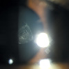
An 11-yr-old boy presented to our clinic because of a gradual decrease of vision in both eyes for 2 months. He had a history of atopic dermatitis, which had been diagnosed at 1 yr old. He had no previous history of ocular trauma. He showed mild eruption and eczema in the face and trunk. He was on follow-up by a local dermatologist, and ophthalmologist. According to the local dermatologist, the skin lesion was stationary and there was no evidence of aggravation of atopic dermatitis. According to the previous medical record, which was obtained from the local ophthalmologist, his visual acuity was 20/20 one year before and both lenses were clear. On examination in our clinic, the best corrected visual acuity was 20/200 in both eyes. Fundus examination was done, and there were no specific abnormalities found. The intraocular pressure was within normal range. The patient showed posterior subcapsular and posterior capsular opacity of the lens in both eyes (Fig. 1). According to the patient's mother, the child had been on herbal medication for 8 months and she could not find any specific change in the child's physical condition during the period. The authors reviewed the prescription of the herbal medication and found that the main composition of the herbal medication was Scutellaria baicalensis, Sophora flavescens and Glycyrrhiza uralensis. The authors also had a full review of the patient's medical record and confirmed that the patient had never taken systemic steroid. We were able to re-confirm that no steroids were added to the herbal medication by reviewing the prescription of the herbal medication.
The patient had a successful cataract extraction under general anesthesia in both eyes at a 1-week interval. Phacoemulsification, posterior chamber intraocular lens implantation, and posterior continuous curvilinear capsulectomy in both eyes were performed. After irrigation and aspiration procedure, a thick posterior capsular opacity located near the optical center of both eyes was observed under the operation microscope, and it was definite that posterior continuous curvilinear capsulectomy was necessary for the recovery of the best visual acuity.
After 3 months postoperatively, the best corrected visual acuity was recovered to 20/20 in both eyes, with a refraction of plano -0.50×150 in the right eye and -0.25 -1.00×170 in the left eye. There was no specific complication after the operation.
DISCUSSION
Atopic dermatitis can be associated with various ocular pathologies, such as cataract, keratoconjunctivitis, keratoconus and retinal detachment (4). There has been a report that patients with facial atopic dermatitis, contact lenses, or both, may rub their eyes more frequently than those with lesions on other parts of the body, increasing their risk of cataract progression (5). The location of the lens opacity differs between regions and reports on this subject. However, anterior or posterior subcapsular opacity holds a major proportion of the cataract types related to atopic dermatitis (6-9). Chen et al. reported a case of a 16-yr-old boy with atopic dermatitis who abruptly developed cataracts in both eyes, while suffering from severe skin itching that had begun 2 months before the initial examination, postulating the retinal peroxidation might play a key role in cataractogenesis (10). The incidence of cataract also has an association with the duration of the steroid therapy and can differ from patient to patient by individual susceptibility (2, 3, 11).
As a rule, the type of cataract opacity, which is associated with atopic dermatitis, is posterior or anterior subcapsular opacity (6-9). However, the manifestation of the opacity type in our patient was posterior capsular opacity, which differs from the anterior subcapsular or posterior subcapsular opacity. The authors suggest that the herbal medication itself seems to give an effect in the development of the cataract in this case.
Herbal medication is a mixed compound medication and it is prescribed by oriental medical doctors according to the patient's age, sex, medical condition, personal constitution, and doctor's personal experience. However, the composition is obscure, and it is difficult to anticipate the effect of the medication. It was seemingly difficult to find out the exact composition of the medication that the patient had taken, perhaps suggesting the ambiguity of treatment with oriental medicine.
Christensen reported that patients who are receiving treatment with atopic dermatitis do not routinely need an ophthalmologic screening test (12). Conversely, Norris and Rivers insist that topical cortocosteroids do not increase the risk of cataract formation. However, they did report a case of a cataract in a severe atopic dermatitis patient who had a high dose of topical and systemic steroid therapy. They also insist that a screening test may be required for the group of patients who are refractive to medical therapy for atopic dermatitis (13).
The finding from this case supports the view that if the patient is taking any kind of medication for a long period (including herbal medication or corticosteroids) ophthalmologic screening tests are highly recommended. There are a lot of hypotheses regarding the cause of cataract formation in atopic dermatitis patients. Stress is thought to induce the accumulation of major basic proteins in the lens epithelial cells. However, in patients with atopic dermatitis, the recovering capacity from this stress may be defective, leading to lens opacity. Elevated serum histamine concentration and rubbing of the eye by the patient can induce the break up of blood-aqueous barrier that can cause the leakage of the self-antigen to the anterior chamber, which causes cataract formation (14). In this case, the cataract showed thick posterior capsular opacity, which differs from the previously known hypothesis. Therefore, this study suggests that there may be some complex mechanism in this case, which is thought to result from the combination of the patient's individual susceptibility and herbal medication composition.
In this case, there are some possibilities that the cataract can be a congenital origin or developed directly due to herbal medications. However, reminding the fact that this patient did not showed clinical symptoms like epiphora, nystagmus, or knitting of the brow, which are often seen in congenital cataract patients, the possibility that the cataract can be a congenital origin is less likely in this case.
Herbal medication is used by many patients suffering from a chronic illness, particularly within Asia. Although there are some positive reports about the efficacy of herbal medication in atopic dermatitis, herbal medication in itself has been shown to cause certain ocular pathologic process such as cataract formation, as it was in our case. Lastly, this report also illustrates the multiple pathogenesis of cataract formation.
 XML Download