

 PDF
PDF ePub
ePub Citation
 Print
Print


INTRODUCTION
Transitional cell carcinoma (TCC) of the upper urinary tract (UUT), including ureteral and renal pelvic TCC, is relatively uncommon. Renal pelvic TCC accounts for 5% of all urothelial tumors, 10% of all renal tumors, and is 3 to 4 times more common than ureteral TCC (1-3). Nephroureterectomy with bladder cuff excision is the standard treatment for UUT-TCC. In general, UUT-TCC shows high local or systemic failure, even after radical surgery (4, 5). T stage, tumor grade, and lymphovascular invasion have been suggested as prognostic factors of UUT-TCC. Of these factors, T stage is the most closely associated risk factor (4-9).
The renal pelvis is surrounded by renal parenchyma and perirenal fat posterolaterally, but is covered with peripelvic fat anteromedially. Despite this structural complexity, all renal pelvic tumors that invade beyond muscularis into peripelvic fat or renal parenchyma are diagnosed as stage pT3 according to the TNM system by the American Joint Committee on Cancer (AJCC) (10). Some urologists and pathologists have been interested in this problematic issue, but few authors have completed studies on substaging pT3 renal pelvic TCC because of their rarity (11, 12).
To elucidate the prognostic impact of peripelvic fat invasion in pT3 renal pelvic TCC, we retrospectively reviewed our single center experience with patients who had been treated for renal pelvic TCC by open surgery.
MATERIALS AND METHODS
The study population
We retrospectively reviewed patients who had been treated surgically for renal pelvic TCC at our institution between 1986 and 2004. Exclusion criteria included the presence of a distant metastasis at diagnosis, unresectable disease, and concomitant invasive bladder cancer. Patients who had concomitant ureteral cancer with a higher T stage than the renal pelvic lesion were also excluded. A study population of 128 consecutive patients was identified with 99 male patients (77.3%) and 29 female patients (22.7%). The median age was 63 yr (30-81) and the median follow-up duration was 56 months (range, 6 to 240).
Treatments and follow-ups
All patients had undergone radical nephroureterectomy, and lymphadenectomy was performed on selected patients when enlarged lymph nodes were encountered intraoperatively or noted on preoperative computed tomography. In this procedure, the hilar and regional nodes adjacent to the ipsilateral great vessel were resected. As postsurgical adjuvant therapy, cisplatin-based chemotherapy was administered to 22 patients. Patient follow-up was relatively uniform including surveillance cystoscopy, urine cytology, abdomen-pelvis computed tomography, and a chest radiography. These tests were performed at 3-month intervals for the initial 2 yr, every 6-months for the subsequent 3 yr, and annually after the initial 5 yr. In addition, whole-body bone scan was checked annually during follow-up.
Study methods
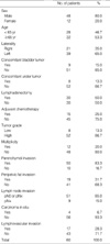
Clinical information was obtained by a retrospective review of medical records in all patients. Survival information was obtained from medical records and our cancer center database. At the last follow-up, 80 patients (62.5%) were alive, 39 patients (30.5%) had died of cancer, and 9 (7.0%) had died of other causes. Pathology slides were reassessed by one urologic pathologist (NHC). Nineteen tumors (14.9%) were stage Ta, 25 tumors (19.5%) were T1, 14 tumors (10.9%) were T2, 60 tumors (46.9%) were T3, and 10 tumors (7.8%) were T4, based on the 2002 American Joint Committee on Cancer (AJCC) TNM staging system (10). Ninety-three tumors (72.6%) were high-grade, and 35 tumors were low-grade (27.4%) according to the 1998 World Health Organization/ International Society of Urologic Pathologists (WHO/ ISUP) classification of papillary urothelial neoplasm (13). Therefore, 60 patients with pT3 disease were eligible for the analyses. The clinical and pathological characteristics of these patients are specified on Table 1.
We investigated the impact of various clinicopathological factors on disease-specific survival. We assessed the following prognostic factors: sex, age, concomitant bladder tumors, concomitant ureter tumors, lymphadenectomy, adjuvant chemotherapy, tumor grade, multiplicity, parenchymal invasion, peripelvic fat invasion, lymph node invasion, carcinoma in situ, and lymphovascular invasion. Kaplan-Meier curves were generated and compared using the log-rank test for the univariate survival analyses. To assess the independent impact of clinicopathological factors on the disease-free survival, Cox proportional hazards regression was used for the multivariate survival analyses. The final result for the multivariate models was obtained using a stepwise forward selection strategy. SPSS for Windows version 12.0 was used for statistical analyses, and a two-sided p value of less than 0.05 was considered to be significant.
RESULTS
Of the 60 patients with pT3 renal pelvic TCC, 30 patients (50.0%) were alive at the final follow-up, and 4 patients (6.7 %) had died of other causes. Twenty-six patients (43.3%) had died of renal pelvic TCC, and the median time to cancer- related death was 28 months (6-102 months). The 5-yr disease-specific survival rate was 59.0%, and the 10-yr disease- specific survival rate was 38.8%.
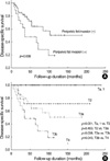
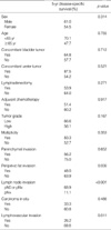
According to the univariate analysis, sex, age, concomitant bladder tumors, concomitant ureter tumors, lymphadenectomy, adjuvant chemotherapy, tumor grade, multiplicity, parenchymal invasion and carcinoma in situ did not influence disease-specific survival. On the other hand, peripelvic fat invasion (p=0.038), lymph node invasion (p<0.001), and lymphovascular invasion (p=0.017) were each significantly associated with disease-specific survival (Fig. 1) (Table 2).
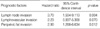
Statistically proven prognostic factors including peripelvic fat invasion, lymph node invasion, and lymphovascular invasion were subjected to multivariate Cox proportional hazards regression analysis to assess the independent impact of these factors. Using this multivariate analysis, significant differences in disease-specific survival rates were found in patients with peripelvic fat invasion (p=0.012) and lymph node invasion (p=0.004). The hazard ratios of disease-specific survival were 2.90 for cases with peripelvic fat invasion and 3.70 in cases with lymph node invasion. However, cases with lymphovascular invasion did not show a significant difference through multivariate analysis (Table 3).
Based on these results, we subdivided the conventional pT3 stage patients into those with peripelvic fat invasion and those without. The prognosis of those with peripelvic fat invasion was more similar to that of pT4 tumors than conventional pT3 tumors, but a statistical difference still existed between them (p=0.043). The prognosis of those without peripelvic fat invasion was similar to that of pT2 disease, and we could not find any difference in the prognosis of patients with pT2 tumors and pT3 disease without peripelvic fat invasion (p=0.453) (Fig. 1).
DISCUSSION
The current staging system of UUT-TCC has been challenged for many reasons. The pathologic findings, pathogenesis, clinical manifestation, and natural history of renal pelvic and ureteral TCC are similar, and thus they have been considered identical diseases. However, questions remain as to whether renal pelvic and ureteral TCC are similar in terms of prognosis because of their apparent anatomical differences. Moreover, several studies have shown that ureteral TCC is associated with a higher local or distant failure rate than renal pelvic TCC and that ureteral TCC is associated with a poorer prognosis (7, 14, 15). The ureter is surrounded by an extensive plexus of ureteral blood vessels and lymphatics, while on the other hand, the renal pelvis is more firmly covered by adjacent tissue, such as the kidney and perihilar adipose tissue. Park et al. suggested that this anatomical weakness of the ureter was responsible for their study results (7).
Another problematic issue has been raised regarding renal pelvic TCC. The staging of renal pelvic TCC has frequently been discussed in the literature (11, 12, 16, 17). In 1971, Grabstald et al. classified 70 cases of renal pelvic tumors into 4 staging groups based on a previous staging of transitional cell carcinoma of the bladder (16, 18). In this study, group IV tumors were defined as those with peripelvic or perirenal fat extensions and adjacent structural invasion. In other words, those patients with peripelvic fat invasion (group IV) were regarded as having higher stage tumors than those with renal parenchymal invasion (group III) (16). Rubenstein et al. modified the Grabstald classification and also separated tumors with peripelvic fat invasion (stage C) from tumors with renal parenchymal extension (stage B) (17). Unfortunately, these series of studies on renal pelvic cancers were small, and although survival percentages for each stage were given, the differences between these stages could not be analyzed statistically. Nonetheless, the AJCC TNM system for UUT-TCC was formulated to standardize staging in 1988, but renal pelvic TCC with renal parenchymal or peripelvic fat invasion were both considered to be identical pT3 stages of renal pelvic TCC (19).
Two previous reports have statistically evaluated the prognostic impact of peripelvic fat invasion. Yoshimura et al. demonstrated that the 3-yr cause-specific survival rates were 55.6% in patients with pT3 renal pelvic tumors containing peripelvic fat invasion and 64.9% in patients without this invasion (12). However, no statistical difference was found between the two groups. A study by Olgac et al. also reported that cases with hilar fat invasion and renal parenchymal invasion had similar outcomes (11). By contrast, in our study, the disease-specific survival rate of pT3 renal pelvic TCC showed a significant difference according to peripelvic fat invasion and lymph node invasion. The prognosis of pT3 tumors with peripelvic fat invasion was more similar to that of pT4 tumors than conventional pT3 tumors. Because of this adjustment, the survival curve of those without peripelvic fat invasion was more similar to pT2 disease. In other words, disease risk has been overestimated in a subset of pT3 renal pelvic TCC patients.
Our results could be explained by a number of mechanisms. The anteriomedial portion of the renal pelvis is surrounded by an extensive plexus of ureteral blood vessels and lymphatics similar to the ureter. This led to the hypothesis that it is easier for those with peripelvic fat invasion than those without to invade lymph vessels. Another explanation may be a problem related to surgical techniques. Because some extent of hilar dissection is required for vascular control, a part of the peripelvic fat layer might be destroyed during this procedure. For this reason, a more meticulous hilar dissection should be required in renal pelvic TCC, especially in the tumors that are located in the anteriomedial part of the renal pelvis. Although adjuvant cisplatin-based chemotherapy did not improve oncological outcomes in the pT3 patients of our study (Table 2), more aggressive adjuvant chemotherapy should be considered for those with peripelvic fat invasion. Outcomes of this strategy, such as whether adjuvant chemotherapy has survival benefits for these patients, should be evaluated based on a larger study population.
There are some limitations in our current study. To clarify risk factors in pT3 patients, we excluded tumors of other stages (pTa, pT1, pT2 and pT4) for the main analysis. In addition to their rarity, this exclusion criterion also caused a relatively small study population. Another limitation is that we did not perform routine lymphadenectomies. Of the 60 patients with pT3 renal pelvic cancers, the rate of lymph node positive pT3 tumors was 15%, though there is a possibility that this rate may be underestimated. Meanwhile, we classified patients with pN0 or pNx as the same entity during our univariate analysis because we could not identify any prognostic differences between them (data not shown). However, this factor might play a role as a confounding factor. Thus, a more refined study population through routine lymphadenectomy would be helpful for controlling these confounding variables, and a larger number of cases would be required to elucidate these observations.
In conclusion, peripelvic fat invasion is a strong prognostic factor in pT3 renal pelvic TCC. More meticulous hilar dissections would be helpful in the surgical treatment of renal pelvic TCC, especially for the tumors located in the anteriomedial region of the renal pelvis. In addition, aggressive systemic chemotherapy should be considered in the presence of peripelvic fat invasion, even if the lymph nodes are not involved.
 XML Download