

 PDF
PDF ePub
ePub Citation
 Print
Print


INTRODUCTION
A mitochondrion is a membrane-enclosed intracellular organelle in most eukaryotic cells. Mitochondria generate most of the cell's supply of adenosine triphosphate (ATP), used as a source of chemical energy. In addition to supplying cellular energy, mitochondria are involved in cell signaling, cellular differentiation, apoptosis, as well as the control of the cell cycle and cell growth. The number of mitochondria in a cell varies widely by organism and tissue type (1). Human mitochondrial DNA (mtDNA) consists of 16,569 base pairs in a supercoiled, double stranded circular molecule. The maternally inherited genome contains 37 genes coding for 13 polypeptides of the mitochondrial electron respiratory chain, 22 tRNAs and two rRNAs necessary for synthesis of the polypeptides (2). Nucleated animal cells contain about 1,000 mitochondria and each mitochondrion contains 2-10 mtDNAs, therefore, a typical differentiated cell has about 3,000 copies of mtDNA for every diploid copy of the genome in the nucleus, non-nucleated cells like platelets and red blood cells have 1-3 and 0 mitochondria per cell, respectively (1).
mtDNA is replicated with a high mutation rate because mtDNA has no protective histones and also lacks an effective DNA repair system, moreover, mtDNA is located near the inner mitochondrial membrane, where it is exposed to oxygen free radicals generated by the respiratory chain (3). Mitochondrial defects occur in a wide variety of degenerative diseases, aging, and cancer. Mitochondrial diseases encompass an extraordinary assemblage of clinical problems, commonly involving tissues that have high energy requirements such as heart, muscle, and the renal and endocrine systems (4). Pathogenic mtDNA defects (deletions and point mutations) affect at least 1 in 15,000 of the adult population (5). To date, over 100 point mutations, 200 deletions and rearrangements have been associated with disease, and new mutations are being described every year (1).
Aplastic anemia (AA) has been traditionally but unhelpfully characterized as 'heterogenous' based on the many different putative etiologies such as idiopathic, secondary causes (radiation, drug and chemicals, viruses and immune diseases), paroxysmal nocturnal hemoglobinuria and pregnancy (6). Among these etiologies, radiation, some drugs and chemicals apparently give rise to mtDNA abnormalities (7-9). We hypothesized that AA may be associated with mtDNA aberrations. In the present study we analyzed the entire mtDNA nucleotide sequences from nine and eight bone marrow (BM) specimens from Korean patients with AA and healthy individuals, respectively. This is the first comprehensive investigation of entire mtDNA genome in patients with AA.
MATERIALS AND METHODS
Patients and healthy subjects
The study included 9 Korean patients with AA (male 5 and female 4) and 8 normal healthy controls (male 6 and female 2). There is no statistical significant difference of age distribution between patients (mean±SD, 39.7±18.7) and normal controls (41.9±14.2) (p=0.395). BM and buccal mucosa (M) specimens were collected after informed consent was obtained following to protocols from the Institutional Review Board of the Chonnam National University Hwasun Hospital (Hwasun, Korea). AA was diagnosed by peripheral blood and BM findings according to criteria of the International Study of AA and Agranulocytosis.
Entire mitochondrial DNA, oligonucleotide primers and direct sequencing
Total DNA was extracted using an AccuPrep Genomic DNA Extraction Kit (Bioneer, Daejon, Korea). Extracted DNA was resuspended in Tris-EDTA (TE) buffer (pH 7.5) containing 10 mM Tris and 1 mM ethylenediamine tetraacetic acid (EDTA). For the direct sequencing of the entire mtDNA genome, we used 20 primer pairs based on a modification of a published protocol in order to obtain 20 partially overlapping segments (10). Each amplified mtDNA product was purified using an AccuPrep PCR Purification Kit (Bioneer) and sequenced with a BigDye Terminator v3.1 Ready Reaction Kit (Applied Biosystems, Foster City, CA, U.S.A.) and an ABI Prism 3100 Genetic Analyzer (Applied Biosystems). Forty oligonucleotide primers derived from Levin et al. (11) were used in sequencing the entire mtDNA genome.
Determination of polymorphism and mutation
mtDNA sequences experimentally obtained were compared to the Revised Cambridge Reference Sequence (http://www.mitomap.org) (12) using the Blast2 program (http://www.ncbi.nlm.nih.gov/blast/bl2seq/bl2.htlm) and the database search tool, MitoAnalyzer (http://www.cstl.nist.gov/biotech/strbase/mitoanalyzer.html, 2001), in order to obtain preliminary evidence for polymorphisms, mutations, and translation of amino acids. Results obtained from AA patients also were compared with those obtained from our concurrently tested normal healthy subjects and established published and unpublished database of mtDNA polymorphism (http://www.mitomap.org), and we treated mtDNA aberrations found in both aforementioned polymorphism database and normal healthy controls as polymorphisms. When mtDNA nucleotide change was only observed in AA patients, it was considered a new mutation. New mutations were confirmed by re-amplification of the region using a separate cell specimen for DNA extraction.
RESULTS
mtDNA nucleotide changes in healthy subjects
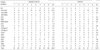
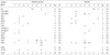
Among hematological normal control subjects, in each controls there were numerous mtDNA sequence variants (No=102, mean=12.8), which consisted of 75 polymorphisms already listed in a published polymorphism database (http://infinity.gen.emory.edu/mitomap.html) and 27 new sequence variations (mean=3.4) not previously recorded, including unpublished mtDNA polymorphisms. These nucleotide changes among the eight normal controls were distributed throughout the mitochondrial genome, and none of new sequence variants led to amino acid changes (Table 3).
mtDNA nucleotide aberrations in patients
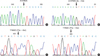
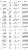
A total of 230 mtDNA nucleotide changes were detected throughout the mtDNA genome. Thirty-two mtDNA variants were newly detected in BM specimens from nine AA patients (mean=3.6). These were 30 substitutions and two deletions at the protein genes for nicotinamide adenine dinucleotide (NADH) dehydrogenase (N=17), cytochrome c oxidase (N=6), cytochrome b (N=3), non-coding region (N=3), ATPase 6 (N=1), and tRNA (N=1) (Table 3). Among them, twelve mutations (38%) harbored amino acid changes (Table 3). There was no predominant mutation site (hot spot). To determine heteroplasmy, we additionally tested buccal mucosa cells. In two of the nine patients there were heteroplasmic sequence variants, which were only present in BM samples, all the other newly detected mtDNA aberrations appeared homoplasmic mutations (Table 1 and Fig. 1).
The pattern of mtDNA sequence variations
Nucleotide substitution rather than deletion accounted for the majority pattern of nucleotide changes. The transition type was more prevalent than the transversion type and deletion. A strong strand bias is more expressed in the HV1 region where transitions between pyrimidines constitute a majority of the mutations, and the frequencies of transversion were higher in the HV1 region than the HV2 region.
Statistical comparisons
Compared with the 2001 Revised Cambridge Reference Sequence and with sequences from normal subjects and patients, we excluded nucleotide changes which found in normal controls and patients. The mean number of mtDNA aberrations in patients with AA (mean=25.6) was higherly than those of healthy individuals (mean=12.8), so a statistical significant difference was noticed (p=0.019) (Table 2). Among the newly detected mtDNA aberrations in patients and healthy subjects, those from healthy subjects did not have amino acid changes (Table 3).
DISCUSSION
mtDNA has been widely used for forensic identification and in anthropological studies because of its abundance and inherent variability. Furthermore, several hundred human diseases have been associated with maternally inherited specific deletions and mutations (13). Somatically acquired mtDNA mutations also have been linked to aging and degenerative diseases, cancer, and autoimmunity (4). A large deletion of mtDNA is a hallmark of Pearson's syndrome, a constitutional disorder that includes sideroblastic anemia (14). mtDNA mutations recently were reported also in apparently acquired sideroblastic anemia and in myelodysplastic syndromes in general (15). While we were unable to confirm these results by amplification and direct sequencing of the entire mtDNA genome in patients and normal controls, we coincidentally observed marked sequence variation in marrow mtDNA among different normal donors (16).
The pathophysiology of acquired AA is complicated. Complexity can be inferred from the diversity of putative etiologic associations from drugs and chemicals to viruses and pregnancy (6). Normal hematopoiesis is dependent upon the transcription and translation of mtDNA into functional proteins. Mitochondrial protein synthesis is selectively blocked by nitrobenzene antibiotic chloramphenicol via its direct action on the large ribosomal subunit of organelle. Acquired deletions of mtDNA in the hematopoietic compartment have also been observed to occur in association with severe pancytopenia and reticulocytopenia (7). For these reasons, in current study we undertook to determine the entire sequence of BM mtDNA from nine AA patients and a comparable number of normal controls. In this study, we found a large number of polymorphisms as well as apparent new mutations in both patients and controls. mtDNA nucleotide changes that were present in our normal controls as well as those in published databases were counted as polymorphisms. Overall, there was significant increase in the number of mtDNA genes harboring polymorphisms or 'new' mutations between AA patients and normal controls. Among the nine patients, twelve mutations producing nonsynonymous alterations were found in NADH dehydrogenase (ND)1, cytochrome c oxidase (CO)I, COII, COIII, ND4, ND5 and cytochrome b. We did not observe small and large deletions that characterize Pearson's syndrome (14), or deletions observed in some other constitutional mtDNA diseases.
The direct relationship between mtDNA mutations and acquired AA development has not studied yet. However, concerning the potential role in mtDNA mutations may contribute to genomic instability (17). For example, deficient mitochondrial ATP production due to mtDNA mutation may promote chromosomal instability. Since the mitotic spindle apparatus depends on ATP-consuming motor proteins, cells with inadequate ATP supply may have difficulty in correctly segregating their chromosomes during mitosis. ATP deficiency may often another pathway to genomic instability through impairment of ATP-dependent DNA repair. Furthermore, respiratory chain dysfunction may impede pyrimidine nucleotide synthesis (17). Thus mtDNA mutations in hematopoietic tissue might be related with AA pathophysiology as a causative and/or aggravating factor. Further study is needed to investigate the direct role of mtDNA mutations of hematopoietic cells in AA pathophysiology and clinical implication.
Most researchers in human mitochondrial genetics feels that heteroplasmic (a mixture of mutant and wild-type mtDNA) is to be considered pathogenic (1). We detected typical heteroplasmic mutations in two patients. It is tempting to associate the limited new mutations seen in these patients with the pathophysiology of AA. Thus, we believe that a more likely and stringent interpretation is that these apparent abnormalities are secondary to the small numbers of stem cell clones operating to support hematopoiesis in this BM failure syndrome.
 XML Download