

 PDF
PDF ePub
ePub Citation
 Print
Print


INTRODUCTION
Reproductive aging is associated with a decline in fertility, which begins in a moderate and steady fashion starting in the third to fourth decade but then accelerates rapidly after age 35 (1). A gradual diminution in the pool of ovarian follicles seems to underlie this decline (2). This age-related decrease in the follicle number and fertility is marked by an increase in follicular-phase serum follicle-stimulating hormone (FSH) levels. This usually starts in women approaching 40 yr of age (3) and is followed several years later by an increase in luteinizing hormone (LH) (4, 5). In addition, changes in estradiol (E2) levels have been described in women with advanced reproductive age (5).
Various endocrinological markers have been used to assess the ovarian reserve, and the accurate and reliable determinations of serum ovarian aging marker levels are essential for safe and successful treatment (6). Previous studies have suggested that anti-Müllerian hormone (AMH) and inhibin B play important roles as ovarian aging markers (7). AMH is secreted by granulosa cells of ovarian follicles and appears to regulate the early follicular development (8). According to a previous study, serum AMH levels decrease with increasing age (9). On the other hand, inhibin B is produced by granulosa cells which characteristically suppress the synthesis and secretion of FSH (10). In addition to its role as a FSH modulator, inhibin B appears to regulate the follicular development and promote follicular growth (11).
Other putative intraovarian regulators of follicle growth include the insulin-like growth factor (IGF) family of proteins. The IGF system is one of several growth factor systems that probably serve adjunctive roles in ovarian follicle development (12). Moreover, IGFs may play an important role in the human preovulatory process, and IGF-binding proteins (IGFBPs) may be valuable biochemical markers in the evaluation of oocytes maturity (13). IGF-I in blood either circulates freely or is bound to binding proteins, mainly to IGFBP-3. Furthermore, serum IGFBP-3 levels have been reported to be positively regulated by IGF-I (14). A small number of studies have explored the positive relationships between follicular fluid IGF-I and ovarian reserve and between follicular fluid IGF-I and serum IGF-I (5). Although serum IGF-I levels have been reported to be downregulated in elderly women and to influence ovarian function (16), the serum IGF-I level is not widely used as an ovarian aging marker and its usefulness has not been clearly documented.
The present study was focused on the usefulness of several ovarian age markers in one population sample with respect to age and menopausal status. Even though the decreases of these hormones with age have been reported (7, 9, 16), the data published to date are not comprehensive enough to reach a consensus regarding reference ranges. Moreover, no study has yet been conducted in an Asian population on these hormones. The purposes of this study were to investigate age-dependent and postmenopausal changes in the serum levels of AMH, inhibin B, IGF-I, IGFBP-3, and FSH and to identify which of these markers better reflects the aging process in Korean women. Furthermore, this study provides diagnostic reference ranges of novel age markers, such as AMH, inhibin B, and IGF-I.
MATERIALS AND METHODS
Subjects
A total of 144 women participated in this study. We recruited healthy, ovulatory women aged 20-29 yr (n=48), 30-39 yr (n=33), and 40-49 yr (n=30) and postmenopausal women aged 50-59 yr (n=33). Premenopausal women were required to have regular menstrual cycles (24-35 days), and postmenopausal women were required to have been in physiologic menopause for at least 1 yr. No participant had any evidence of endocrine disorders (normal prolactin and thyroid-stimulating hormone levels, and no evidence of polycystic ovarian syndrome), and all participants were required to have a body mass index (BMI) of 19-26 kg/m2, both ovaries present, and an absence of medical or reproductive disorders (including any history of infertility), and no participant was taking hormone medications.
Hormone assays
Blood samples were obtained by venipuncture on cycle day 3 from regularly menstruating women or at random from postmenopausal women to measure the serum levels of AMH, inhibin B, IGF-I, IGFBP-3, FSH, LH, and E2. Serum was separated from blood samples and stored at -20℃ until assayed. Samples from a given subject were analyzed for each hormone in the same assay to avoid inter-assay variation.
Serum levels of LH and FSH were measured by immunoradiometric assay (IRMA) using commercial kits (Biosource, Nivelles, Belgium). The detection limits of the assay were 0.2 mIU/mL for LH and 0.1 mIU/mL for FSH. Intra- and inter-assay coefficients of variation (CV) were 3.2% and 6.7% for LH, and 3.3% and 7.1% for FSH, respectively. Samples were analyzed for E2 using radioimmunoassay (RIA) using commercial kits (Biosource, Nivelles, Belgium). The detection limit of this assay was 10 pg/mL, and intra- and inter-assay CVs were 4.9% and 5.2%, respectively. Serum levels of IGF-I and IGFBP-3 were determined by IRMA using reagents supplied by Diagnostic Systems Laboratories (DSL Inc., Webster, U.S.A.). Its detection limits were 2.06 ng/mL for IGF-I and 0.5 ng/mL for IGFBP-3, and its intra- and inter-assay CVs were 5.1% and 9.1% for IGF-I, and 5.2% and 7.1% for IGFBP-3, respectively.
Serum levels of AMH and Inhibin B were determined using enzyme-linked immunosorbent assays (ELISA) using commercial kits from Diagnostic Systems Laboratories (DSL Inc., Webster, U.S.A.). The detection limits of this assay were 0.017 ng/mL for AMH and 7 pg/mL for Inhibin B, and its intra- and inter-assay CVs were 4.6% and 8.0% for AMH and 5.6% and 7.6% for inhibin B, respectively.
Statistical analysis
Data were analyzed by age group in premenopausal women. Differences between age groups were tested by one-way analysis of variance (ANOVA), followed by Turkey's multiple comparison test. Comparisons between pre- and postmenopausal women were made using Mann-Whitney U test. The abilities of hormone levels to discriminate between pre- and postmenopause women were investigated using receiver operating characteristic (ROC) analysis. The diagnostic values of the various hormone markers were evaluated by comparing areas under the curves (ROCAUC) (23). Statistical analyses were performed using SPSS 12.0 for Windows (SPSS Inc., Chicago, IL, U.S.A.), and p values of <0.05 were considered statistically significant. We drew fitting plots of hormone levels versus age. Mean data values with error bar are presented in the fitting plots. The x-axis was divided into intervals of 5 yr and each curve was fitted using linear, Gaussian, Lorentzian, and sigmoid functions using Mathematica 5.0 (Wolfram Research Inc., Champaign, IL, U.S.A.)
RESULTS
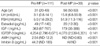
The characteristics of the premenopausal subjects and their serum hormone levels on cycle day 3 are shown in Table 1. Serum levels of AMH, IGF-I, and IGFBP-3 decreased and those of FSH increased significantly with age. Serum levels of E2, LH, and inhibin B showed no significant differences. However, serum LH was higher and inhibin B was lower in women in their 20-30's than in 40's. AMH and IGF-I levels consistently decreased with age, and showed significant changes across all ages, whereas IGFBP-3 and FSH levels showed a significant difference only between women in their 30's and 40's.
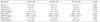
Median ages and serum hormone levels of premenopausal and postmenopausal women are presented in Table 2. Data for AMH and inhibin B were not normally distributed in the postmenopausal women, mainly because the majority of values were below the detection limit. Hence the data were presented as medians and ranges, and a nonparametric method was used for comparison between two groups. Premenopausal and postmenopausal women had median ages of 31 (range 20-49) yr and 56 (range 50-59) yr, respectively. Serum levels of LH, FSH, E2, IGF-I, AMH, and inhibin B showed significant differences between pre- and postmenopausal women (p<0.001). However, serum IGFBP-3 levels did not change significantly after menopause. As expected, LH and FSH levels in postmenopausal women were significantly higher and those of E2, IGF-I, AMH and inhibin B levels were significantly lower than in premenopausal women.
AMH was undetectable in 20 of 33 postmenopausal women, whereas it was only undetectable in 2 of 111 premenopausal women. Inhibin B was undetectable in all postmenopausal women and in 10 of 111 premenopausal women; moreover, these 10 women were older than 40 yr old.
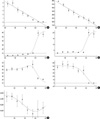
Serum levels of AMH and IGF-I showed linear decreases from the earlier age (Fig. 1A, B), whereas FSH and LH levels showed sigmoid increases and showed greatest increase after menopause (Fig. 1C, D). Serum E2 levels were fitted using sigmoid and Lorentzian functions (Fig. 1E) and inhibin-B levels showed a sigmoid decrease (Fig. 1F), and both of these hormones decreased after menopause. Serum IGFBP-3 levels were fitted using Gaussian function with a minimum value at the boundary between pre- and postmenopause (Fig. 1G).
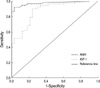
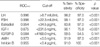
Table 3 shows the results of ROC curve analysis for serum markers. This analysis showed that the diagnostic accuracy of AMH (ROCAUC=0.943) for menopausal status was similar to those of FSH (ROCAUC=0.998), LH (ROCAUC=0.996), and inhibin B (ROCAUC=0.945), and was better than IGF-I (ROCAUC=0.875). Cutoff levels providing desired sensitivities and specificities can be deduced from ROC curves. When the point on a curve closest to the upper left corner of the box corresponding to 100% sensitivity and 100% specificity (0% false positivity) was selected, it resulted in cutoff levels for AMH of <0.46 ng/mL and for inhibin B of <0.4 pg/mL in terms of identifying postmenopausal subjects. The sensitivity and specificity corresponding to these cutoffs were 92% and 97% for AMH, and 91% and 100% for inhibin B, respectively, and the cutoff level of IGF-I was <231.5 ng/mL with a sensitivity and specificity of 90% and 76%, respectively. The sensitivity and specificity of AMH were similar to those of FSH, LH, and inhibin B, and were better than those of IGF-I.
DISCUSSION
Several studies have investigated changes in markers of ovarian follicular reserve. Serum FSH levels increase in old reproductive age women, a fact that has been well documented and recognized for many years by many investigators (5). In women approaching 40 yr of age, serum FSH levels usually begin to rise, which reflects a reduction in the number of early antral follicles present that can be recruited to ovulate (3). Serum FSH levels increase over time because inhibin B and E2 production are reduced by a diminished cohort of growing follicles (18, 19). Nevertheless, prior to age 40, FSH levels are not correlated with age, which confirms the lack of correlation between FSH and age in women aged 20-35 (20, 21). However, the proper assessment of ovarian aging at an early stage is crucial in terms of counseling patients about their possibility of pregnancy, either spontaneously or during fertility therapy. Therefore, a new marker of ovarian aging in younger women is needed.
Previous studies have reported that serum AMH levels are closely related to the early antral follicle count, and moreover, this relationship was found to be remarkably more strong than those of inhibin B, E2, FSH, or LH (8, 9, 22). The serum AMH level decreased with ages in premenopausal women and postmenopausal women, which is in line with the findings of de Vet et al. (7). In the present study, it was found that serum AMH levels in normal ovulatory women reduced with advancing age before changes in other markers (e.g., FSH and inhibin B) were apparent, and that AMH was undetectable in most of postmenopausal women. These results are in line with those of previous studies and suggest that AMH could be used as a novel marker of ovarian aging.
Age-dependent decreases of IGF-I may occur secondary to age-dependent reductions in growth hormone secretion (23, 24). IGF-I has been shown to serve as an intraovarian regulator of follicle function in rodents and to exerts a direct effects on human and rodent granulos a cell function (25, 26). Moreover, IGF-I, in conjunction with gonadotropins, appears to promote follicle growth (27) and steroid secretion (28) and to act as an antiatretic hormone (29). Although serum IGF-I levels have been reported to be attenuated in elderly women (16) and follicular fluid IGF-I levels have been demonstrated to be related to the ovarian reserve (15), serum IGF-I levels are not widely used as an ovarian aging marker and the usefulness of serum IGF-I has not been clearly documented in this context. Based on a high correlation between serum and follicular fluid levels of IGF-I and on the positive correlation between follicular fluid level and an ovarian reserve (15), it can be speculated that serum IGF-I and ovarian reserve are positively correlated. The present study demonstrates age-dependent and postmenopausal changes in the serum levels of IGF-I, which suggests that serum IGF-I is an another candidate marker of ovarian reserve.
The results of the present study indicate that serum levels of AMH, IGF-I, and IGFBP-3 decrease and that those of FSH increase significantly with age in premenopausal women, but serum levels of inhibin B do not decrease significantly with age. In addition, FSH levels in normal ovulatory women in their 20's and 30's were similar, which is in line with previous reports on inhibin B and FSH (7, 20, 21). In contrast, serum levels of AMH and IGF-I were significantly different in women in these age groups. It is noteworthy that serum levels of AMH and IGF-I significantly changed at earlier ages than did those of the other hormones in premenopausal women; moreover, the main limitation of conventional ovarian reserve markers such as FSH, LH, and E2 is that their serum levels change relatively late.
All hormones examined showed significant differences pre- to postmenopause except IGFBP-3. Inhibin B was practically undetectable after menopause, and AMH was undetectable in 20 of the 33 postmenopausal women. This is supposed to be because inhibin B and AMH are produced by the granulosa cells of ovarian follicles (7, 10). Several studies have investigated changes in AMH and inhibin B, and also found that they ultimately become undetectable after menopause (7, 30).
To follow the age-dependent changes of each marker more precisely, we fitted their hormone level versus age plots. After menopause, serum FSH and LH levels increase markedly and E2 and inhibin B levels decrease markedly, but no changes in these markers were observed during earlier premenopausal ages. The fitting curves of the AMH and IGF-I serum levels versus ages showed a linear decrease from the earlier age. The present study more precisely characterized the usefulness of AMH and IGF-I during early reproductive ages. These results suggest that AMH and IGF-I are better markers of ovarian reserve in premenopausal women than other hormone markers, such as FSH, LH, E2, and inhibin B.
To determine whether AMH or IGF-I better reflects the aging process in women, we compared the diagnostic accuracies of hormone levels in terms of differentiating between pre- and postmenopausal status (Fig. 2). ROCAUC analysis showed that the diagnostic accuracy of AMH for menopausal status was similar to those of FSH, LH, E2, and inhibin B, and that it was better than IGF-I. The reason for the inferiority of IGF-I to AMH and other markers appears to be that IGF-I levels decrease continuously after menopause, whereas FSH, LH, and E2 levels are fairly stable and AMH and inhibin B levels vanish after menopause.
In conclusion, the present study provides strong evidences that the serum AMH level is an important marker of reproductive aging in women. It was also found that serum IGF-I is a good candidate ovarian aging marker, especially in premenopausal women. In addition, the results of this study provide useful reference data of serum AMH ranges in Korean populations. Further research of large scale and longitudinal design is necessary to confirm our results.
 XML Download