

 PDF
PDF ePub
ePub Citation
 Print
Print


INTRODUCTION
The management of colorectal cancer, especially that of rectal cancer, has changed in many aspects during the last few decades (1-3). Previous studies based on patients' clinical outcome and hospital data showed that a surgeon's operation volume and the level of the hospital influenced the treatment outcome, and the surgeon's operation volume has been suggested to be the most important contributing factor to the outcome (4-8). The volume-outcome association is more pronounced in rectal cancer than in colon cancer due to the high complexity of rectal surgery (6). Although the difference in surgical procedures has been known to be the most important influencing factor (6), more detailed differences have not been well evaluated. In the present study, therefore, we attempted to unravel the differences of rectal cancer management and then to compare the differences, based on the surgeons and the hospitals.
MATERIALS AND METHODS
Questionnaires describing preoperative evaluation, operative procedure, and postoperative surveillance in 39 major categories were sent out to the members of the 'Korean Society of Coloproctology', including the surgeons at 30 university hospitals, in August 2004. The surgeons who operated rectal cancer in their hospitals were requested to respond to the questionnaires. Sixty responses were received during the three months' period. After the data were coded blindly into the database, the results were compared according to the surgeon's annual operation volume, the level of the hospital, and the surgeon's age. Statistical analyses were performed by chaisquare test using SPSS 11.0® (SPSS Inc., Chicago, IL, U.S.A.).
RESULTS
Respondents
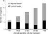
Thirty three respondents (55.0%) operated more than 50 rectal cancer cases (high-volume surgeons), and 37 respondents (61.6%) worked at university hospitals or in other tertiary care facilities (high-level hospitals) (Fig. 1). Twenty respondents (33.3%) were 40-yr old or younger (young surgeons), and the respondents' age was not significantly different according to the surgeon's operation volume or the level of the hospital.
Preoperative evaluation
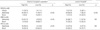
Preoperative rigid sigmoidoscopy was used routinely by 40.0% of the respondents and selectively by 36.6% of the respondents, whereas 23.3% did not use the examination. Colonoscopy, however, was performed routinely by 88.3% of the respondents and selectively by 10.0% of the respondents. The surgeons participated in colonoscopic examination alone or together with endoscopist at 50% of the respondents' hospitals. Only 21.7% of the respondents routinely used barium enema, whereas 16.6% did not use this examination for preoperative evaluation. One respondent had a limitation for usage of magnetic resonance imaging (MRI) and positron emission tomography (PET) during their evaluation, while others performed the examinations without limitation in their hospital or neighboring institution. Liver imaging by computerized tomography (CT) was done by 96.7% of the respondents, and abdominal ultrasound was additionally used by 30% of the respondents. Significant differences were found in endorectal ultrasound (ERUS), pelvic MRI, and 18F-fluorodeoxyglucose-PET: ERUS and MRI were significantly different according to the surgeon's operation volume, and ERUS was also significantly different according to the level of the hospital (Table 1). The selective use of PET was significantly different according to the surgeon's operation volume (Table 1).
Preoperative radiation therapy
Preoperative radiation therapy was given fractionatedly in 4 to 6 weeks with the radiation dose ranging from 4,500 to 5,500 cGy. Forty six respondents (76.7%) performed preoperative radiation therapy, which was significant according to the surgeon's operation volume (90.9% of high-volume surgeons vs. 22.2% of low-volume surgeons), the level of the hospital (89.1% of high-level hospitals vs. 56.5% of low-level hospitals), and the surgeon's age (95% of young surgeons vs. 67.5% of aged surgeons). As for the purpose of preoperative radiation therapy, 80.4% of the 46 surgeons performed it for the purpose of sphincter preservation.
Operation
Total mesorectal excision (TME) was considered as the principle in rectal cancer surgery by 96.6% of the respondents, and 68.9% of them performed it only for mid and low rectal cancer. Inferior mesenteric artery was ligated at its root routinely by 26.6% and selectively by 68.3% of the respondents. Rectal irrigation was performed before dividing below a tumor by 81.6% of the respondents. During an operation, 2 cm of distal mucosal resection margin was considered as safe by 62.0% of the respondents; and 1 cm or less by 29.3% of the respondents. Paraaortic lymph node dissection was performed routinely (5%), selectively (75%), or never (20%); and lateral pelvic lymph node dissection was performed selectively (93.3%) or never (6.6%). Coloanal anastomosis was performed by 88.3% of the respondents, and the most preferred type was straight form (77.4%), J-pouch (15.1%) and coloplasty (7.5%). Diverting stoma was performed routinely (20.8%) or selectively (54.7%) during coloanal anastomosis, however, 24.5% did not fashion diverting stoma during coloanal anastomosis.
Operative procedures were not significantly different according to the surgeon's operation volume, the age, or the level of the hospital.
Postoperative management
Adjuvant chemotherapy was given by surgeons in 73.3% of the respondents' hospitals and by medical oncologists in 15.0% of the respondents' hospitals. In 11.7% of the respondents' hospitals, chemotherapy was given by surgeons or medical oncologists according to the patients. Postoperative radiation therapy was given according to the stage: 21.6% of the respondents routinely for T3N0 and 50.0% for T3N1 or T3N2.
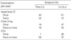
Carcinoembryonic antigen (CEA), abdominopelvic CT, chest radiography, and colonoscopy were the main surveillance tools after operation. There were no significant differences in the use of these tools according to the surgeon's operation volume, the level of the hospital, or the surgeon's age (Table 2). Colonoscopy was performed annually (66%) or biannually (28%) by 95% of the respondents for the fllowing five years after operation. On the other hand, rigid sigmoidoscope was used only by 43.3% of the respondents. While the regular follow-up during the 5 yr postoperative period was not significant, the regularity of follow up after the initial 5 yr was significantly different showing 90% of high-volume surgeons and 62% of low-volume surgeons regularly performed it every one or two years (p<0.05).
DISCUSSION
This study examined the differences in the rectal cancer management by the surgeons in Korea. Rectal cancer has been increasing in Korea, including 5,865 new cases reported in 2002. We strongly believe that more than 4000 operations of rectal cancer cases are performed annually by the surgeons who are included in this study. Therefore, considering resectability (9), it is quite possible that this study covers more than 70% of the rectal cancer operations in Korea.
The previous studies with oncologic outcome defined the minimal volume for the experienced surgeon as one or two operations per month, and the cutoff value of other studies was about 15 cases or less (4-7). On the other hand, the studies with operative technique and outcome defined the cutoff value as 50 or 75 operations per year (10-12). In this study, the high operation volume was defined as more than 50 operations per year, and the majority of the surgeons (75%) operated more than 30 rectal cancer cases per year. It was possible that may surgeons with a low annual volume were not included in this study.
Recent advances in the management of rectal cancer include improved preoperative pelvic imaging, preoperative chemoradiation therapy, and improved rectal dissection (1, 2, 9, 13). In the present study, significant differences have been found in the preoperative evaluation with ERUS, MRI, and PET. ERUS has been used for more than 20 yr and is known to be accurate for the early stage. On the other hand, MRI is more informative for the advanced stage and especially for the mesorectal fascia which is important for keeping safe circumferential resection margin during pelvic dissection (14-17). PET is increasingly used for both local evaluation and distant metastasis (18). In the present study, these preoperative evaluations have been found to be different according to the surgeon's operation volume, yet only ERUS has been found to be significantly different according to the level of the hospital.
There were general agreements among the surgeons on the operative procedure (more than 75% of the respondents agreed): TME as the primary technique in rectal cancer surgery; selective use of paraaortic and pelvic lymph node dissection; rectal irrigation; and coloanal anastomosis for sphincter preservation. On the other hand, the operation of upper rectal cancer was controversial among the surgeons, which was also noted in other studies (1, 19-21). Controversies on diverting stoma for low rectal cancer operation (20, 22, 23) were also noted in this study. Regardless of the general agreements or the controversies, a significant difference was not found according to the surgeon's operation volume, the surgeon's age, or the level of the hospital.
Unlike the operative procedure, preoperative radiation therapy was significantly different according to the surgeon's operation volume, the level of the hospital, and even the surgeon's age, and it was the only significant factor according to the surgeon's age. Postoperative surveillance varied among surgeons and was performed differently according to the patient's condition. Colonoscopy was frequently used rather than sigmoidoscopy because of the comparable expense of these medical examinations in Korea (24). The surveillance system is affected by socioeconomical factors or by insurance systems, which could be different according to the country. These days, intensive surveillance is increasing for the early detection of metastatic or recurrent diseases, and the examinations were found more frequent in this study than in other recommendation (25).
This study was conducted by using a questionnaire, and several limitations were present. The questionnaire was completed by the surgeons themselves and many low-volume surgeons did not reply. All the complexities and the individualized treatments could not be assessed only by this questionnaire. The relationship between the surgeon's actual practice and the clinical outcome could not be proved with this study. Nevertheless, the surgeon's intention and behavior during the treatment of rectal cancer have accurately been evaluated by this study, and the recent trends of rectal cancer operation were also revealed.
In conclusion, preoperative loco-regional evaluation was significantly different according to the surgeon's operation volume, and preoperative radiation therapy was significantly different according to the surgeon's operation volume and the level of the hospital. These results could be considered as the factors to influence volume-outcome relationship in rectal cancer treatment.
 XML Download