

 PDF
PDF ePub
ePub Citation
 Print
Print


INTRODUCTION
Intracranial tuberculoma is a rare condition and is one of the causes of intracranial mass lesions. However, its reported incidence varies, from 0.15% to 30% in intracranial mass lesions (1, 2). Intracranial tuberculoma is a serious and fatal disease, but its frequency has increased in recent years due to its occurrence in immune-compromised hosts (3). The diagnosis of intracranial tuberculoma is often difficult because conventional magnetic resonance (MR) imaging of tuberculoma reveals various findings. In addition, it requires differential diagnosis with metastatic tumors, abscesses, malignant brain tumors, and other brain lesions that can be associated with acute myeloid leukemia (AML) (4). Moreover, neurological symptoms are not typical (5).
We describe an interesting case of intracranial tuberculoma that subsided after anti-tuberculous medication in a patient with AML.
CASE REPORT
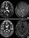
A 52-yr-old man visited our hospital and was diagnosed with AML in June 2006. Bone marrow aspiration revealed 75% blasts, and the French American British classification was M2. Cytogenetic study showed normal chromosomes, and the patient was classified into the intermediate-risk group. Treatment with idarubicine (12 mg/m2 day 1-day 3) and Ara-C (100 mg/m2, day 1-day 7) remission induction chemotherapy was initiated. During induction chemotherapy, the patient experienced a neutropenic fever, and empirical antibiotics were administered to treat anal abscesses. After the first course of induction chemotherapy, the patient achieved complete remission, and we treated him with three times high-dose Ara-C (2 g/m2 Bid, days 1, 3, 5) consolidation chemotherapy sequentially. After the third cycle consolidation treatment, he visited the outpatient department because of right-side paresthesia in November 2006. The complete blood count showed WBC 4,200/mL (normal, 4,800-10,800), Hgb 11.3 g/dL (normal, 12-18), and platelet 160,000/mL (normal, 130,000-450,000), and a leukemic relapse was not suspected. For further evaluation of neurologic symptoms, MR images of the brain were checked. The gadolinium-enhanced brain MR imaging showed multiple ring-enhanced lesions with mild perilesional edema in the brain; the lesions were located in the cerebrum, cerebellar hemispheres, and pons. They were mostly located at the gray-white matter junction (Fig. 1). For differential diagnosis of granulocytic sarcomas, a bone marrow biopsy was performed. On the bone marrow biopsy, focal abnormal cell clusters were observed on the paratrabecular area, and showed a non-remission state. Cerebrospinal fluid (CSF) analysis showed: pH 7.799, WBC negative, protein 100 mg/dL (normal, 15-50), glucose 49 mg/dL (normal, 40-70), and adenosine deaminase 28.8 IU/L (normal, 0-8). Mycobacterium tuberculosis polymerase chain reaction (PCR) and India ink staining showed negative findings.
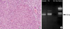
To confirm diagnosis, a stereotactic brain biopsy was performed, but only reactive gliosis was observed. An open brain biopsy was then conducted. The gross finding showed a mass with a central necrotic portion surrounded by hard wall. The histopathological findings demonstrated chronic granulomatous inflammation with tuberculous-PCR positivity, but acid-fast bacilli staining was negative (Fig. 2). A routine chest radiograph showed non specific findings. High-resolution computed tomography (HRCT) was assessed because of the possibility of pulmonary tuberculosis. However, HRCT showed no abnormalities. The patient was treated with isoniazid, rifampin, ethambutol, pyrazinamide, and high-dose steroid. After treatment for 1 week, paresthesia improved. A follow-up brain MR imaging after one month showed decreased size and number of ring-enhanced lesions and improving perilesional edema. Therefore, we planned for anti-tuberculosis maintenance treatment to be prolonged for about 9 months as well as additional reinduction chemotherapy.
DISCUSSION
Mortality and morbidity in patients with acute leukemia is mainly related to relapse or infection. The cellular immunity is extremely disrupted following high-dose chemotherapy. Opportunistic infections from bacterial, viral, and fungal pathogens can occur at this time (6). In areas where tuberculosis is endemic, the general population is exposed to the tubercle bacillus throughout life. The dysregulation of immune T-cells would render these patients immunologically unable to defend themselves against Mycobacterium tuberculosis. The incidence of microbiologically tuberculous infection in acute leukemia was reported about 2.2%, and the most common site of tuberculous infection was the lung (6). The intracranial tuberculomas in acute leukemia was not a common infection site. Only 1% of immune-competent patients with tuberculosis develop an intracranial tuberculoma, usually as part of miliary tuberculosis (7-9).
The diagnosis of intracranial tuberculomas is difficult to make in immune-compromised patients, as misleading results may arise from non specific symptoms (e.g., headache, fever, weight loss, and weakness) (10). It is especially difficult to distinguish symptoms of a brain tumor from those of an abscess (e.g., seizure, headache, visual disturbance, and hemiparesis) (1, 11), and diagnosis of intracranial tuberculomas based on radiographic and bacteriologic investigations is also quite difficult (3, 12). Cultures of CSF of tuberculoma are usually found to be negative (5), but positive PCR results in the biopsied tissue are found in 60% to 80% of cases. In most cases of intracranial tuberculomas, the initial clinical diagnosis was a neoplasm. For this reason, clinicians must always be aware of neoplasm, and should make the differential diagnosis from various tumorous conditions in the brain (7, 10).
An association with pulmonary tuberculosis was reported from 25% to 83% of cases, and 40% of patients had a history of tuberculosis (1, 2, 5, 11). Therefore, the patient history and findings on chest radiology should be taken together in the diagnosis of intracranial tuberculomas.
Conventional MR imaging is very useful to detect small lesions and to distinguish tuberculomas from other inflammatory lesions or brain tumors. A slightly hyperintense rim with perilesional edema on T1-weighted images was also described in pyogenic abscesses, and these findings are similar to those of tuberculomas. However, the hypointensity or isointensity that is frequently seen in the central portions of tuberculomas on T2-weighted images can be used to differentiate tuberculomas from pyogenic abscesses (5). Diffusion-weighted imaging of the brain showed homogeneous high signals in tuberculomas. In our case, MR imaging of the brain showed a similar finding of tuberculomas, but multiple brain lesions and patient disease status (non-remission state) were needed for differential diagnosis between tuberculomas to other brain tumors (metastatic tumors or granulocytic sarcomas) (4, 13, 14).
Many brain lesions could be associated with myeloid leukemia, and the diagnosis was difficult to establish in this case because of diffuse multiple parenchymal lesions mimicking granulocytic sarcomas or abscesses (15). The case described in this report was an interesting case of intracranial tuberculosis in AML.
 XML Download