

 PDF
PDF ePub
ePub Citation
 Print
Print


INTRODUCTION
Traumatic tricuspid regurgitation has been thought to be a rare complication of blunt chest trauma, although its reported frequency has increased over the past four decades (1). Various kinds of trauma can cause traumatic valvular injury such as motor vehicle accidents (1-4), horse kicks (5), cardiopulmonary resuscitation (6), and falls (7). To our knowledge, however, this is the first report of traumatic tricuspid regurgitation after internal cardiac massage.
In this case, external and internal cardiac massage were performed due to cardiac arrest in a patient who underwent offpump coronary artery bypass surgery (OPCAB). After successful cardiopulmonary resuscitation, the patient was transferred to the operating room to evaluate the hemodynamic instability and close the chest. During surgical preparation, a transesophageal echocardiography revealed severe tricuspid regurgitation due to ruptured papillary muscle. The papillary muscle was repaired under emergency cardiopulmonary bypass, and there was no remaining tricuspid regurgitation.
CASE REPORT
A 66-yr-old man with a 30-yr history of hypertension presented with episodes of chest pain over 10 days. His blood pressure had been controlled with atenolol. A treadmill test showed ST segment depression at stage 2 for 4 min. The coronary angiography at admission revealed 40%, 80%, and 50% luminal narrowing of the left main, the left anterior descending, and the left circumflex artery, respectively. OPCAB was undertaken via median sternotomy. Transesophageal echocardiography performed after the induction of anesthesia revealed left ventricular hypertrophy, normal contractility (left ventricular ejection fraction, 60%), and no abnormal findings in valves. The right internal mammary artery was anastomosed to the left anterior descending artery, the left internal mammary artery was anastomosed to the diagonal branch, and a saphenous vein graft was interposed between the aorta and the obtuse marginal branch.
During the anastomoses, there were no abrupt changes in hemodynamic parameters, and there was no difference in cardiac contractility or valvular function between preoperative and immediate postoperative echocardiographic findings. Five hours after admission to the intensive care unit (ICU), the patient was alert, and the recovery of respiratory muscle power was thought to be enough for spontaneous breathing. At this time, his blood pressure was 102/59 mmHg and central venous pressure was 8-9 mmHg. Therefore, the patient's position was changed from supine to sitting, and the ventilator rate was reduced from 10/min to 8/min in an attempt to wean the patient from the ventilator. After that, the pa tient began to perspire heavily, and his systolic blood pressure decreased from 120 mmHg to 60 mmHg. After returning to supine, the patient became unconscious, and radial artery pressure showed flat wave with 18 mmHg of central venous pressure. External cardiac massage was performed for 15 min. Atropine 0.5 mg and epinephrine 1.0 mg were injected twice intravenously. Electrocardiogram showed ventricular fibrillation, which was resistant to serial monophasic defibrillation of 200, 300, and 360 J. Therefore, the median sternotomy wound was opened to initiate internal cardiac massage. When the sternum was opened, the heart was collapsed and seemed to be grossly normal. Internal cardiac massage was continued until a normal sinus rhythm was restored after the direct cardioversions at 20 and 30 J. Severe hypovolemia was suspected, due to the low diastolic pulmonary pressure and central venous pressure as well as the empty and small heart. Immediately after the sinus rhythm was restored, the laceration and contusion on the right ventricle was observed. Since the laceration was shallow and there was no active bleeding, it was directly sutured in ICU using 5-0 monofilament polypropylene (Surgipro II, Tyco healthcare Group, Mansfield, MA, U.S.A.). There was no progress in the lesion on the right ventricle. However, after resuscitation, high doses of vasopressors (norepinephrine 0.1 µg/kg/min, vasopressin 8 unit/hr, and epinephrine 0.5 µg/kg/min) were required to maintain the blood pressure, and the diastolic pulmonary arterial pressure and central venous pressure were consistently increased, although the cardiac rhythm was stable. The surgical team decided to fully evaluate the heart and close the sternum in the operating room, and thus, the patient was transferred into the operating room.
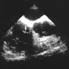
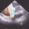
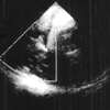
At the operating room, the patient's heart rhythm was sinus of 57/min, systemic blood pressure was 100/60 mmHg, central venous pressure was 16 mmHg, pulmonary artery pressure was 45/23 mmHg, and the cardiac index was 2.1 L/min/m2. Right heart dysfunction was suspected after inspection of the dilated right ventricle, and a transesophageal echocardiographic examination was performed to evaluate the reason for hemodynamic instability. Avulsion of the anterior papillary muscle and cordae (Fig. 1) and severe tricuspid regurgitation were found (Fig. 2). The right ventricle was markedly distended, and the contractility of the right ventricular free wall and the global left ventricular contractility were reduced without mitral regurgitation. The severity of tricuspid regurgitation was grade III-IV/IV. The ruptured papillary muscle was fixed to the right ventricular wall under cardiopulmonary bypass. Measurements of coronary blood flow through all bypass grafts were within normal ranges and additional coronary bypass grafts did not seem to be needed. Right and left ventricular contractility were depressed postoperatively, although the tricuspid regurgitation disappeared. An intraaortic balloon pump (IABP) was inserted immediately after the surgery because of low blood pressure and cardiac output. After that, it was possible to decrease the dose of inotropics and vasopressors. The IABP was removed two days after the surgery. The patient was discharged 14 days after surgery (Fig. 3).
DISCUSSION
Traumatic tricuspid insufficiency is rare but well described, with various symptoms (8-10). Traumatic cardiac injuries are often overlooked because of the inexperience of the physician or due to serious compounding injuries that may obscure the cardiac injury (3). However, traumatic cardiac lesions have been reported with an increasing frequency because of the increased awareness of the relationship between chest trauma and cardiac lesions and the wide application of echocardiography during the past two decades (2). Although many kinds of chest injuries are known to be related to valve injuries (1-7), this is the first report of valve injury after internal cardiac massage to the best of our knowledge.
The mechanism of the valvular injury during blunt chest trauma is thought to involve an abrupt elevation of ventricular pressure from a sudden compression of the heart when the atrioventricular valve is closed (2, 10). The same mechanism may be applied to this case, or a papillary muscle rupture due to a preexisting myocardial ischemia could be a possible mechanism. However, since the heart was grossly normal when the sternum was opened during OPCAB and right coronary artery blood flow was intact, the possibilities of these explanations are fairly low. By contrast, following two facts might strongly support that the tricuspid regurgitation was associated with internal cardiac massage in this case. Firstly, a newly developed laceration and contusion on the right ventricle was observed after internal cardiac massage. Secondly, the surgeon confirmed that the lesion corresponded to the position of the anterior papillary muscle of tricuspid valve in the surgical field. Kinking of the chordae or friction with intracardiac structures from direct manipulation of the heart during the surgery or internal cardiac massage can cause tension to break papillary muscle and result in direct valvular trauma. Another possible mechanism is an interaction between the tricuspid valve and pulmonary artery catheter. Direct contact of pulmonary artery and valve tissue can cause valvular injury during internal cardiac massage.
Jahnke et al. (10) classified traumatic tricuspid injury into three types: rupture of the papillary muscle, chordal rupture, and tear in the leaflet. The reported frequency of each type of injury is highest for chordal rupture, followed by rupture of the papillary muscle and leaflet tear (2, 11, 12). Chordal rupture is associated with a more insidious history extending from 10 to 25 yr, whereas papillary muscle rupture becomes symptomatic earlier. van Son et al. (2) suggested that early diagnosis and surgical treatment could prevent progressive right heart failure and maintain a normal sinus rhythm.
In this case, biventricular dysfunction was suspected due to systemic hypotension, low cardiac index, and elevated central venous pressure and pulmonary artery pressure after open chest cardiopulmonary resuscitation. Coronary blood flow interruption was suspected as a reason for hemodynamic instability, and cardiac contusion due to the internal cardiac massage was thought to be the cause of right ventricular dysfunction because there was no right coronary lesion preoperatively. However, transesophageal echocardiography for cardiac function assessment incidentally revealed tricuspid regurgitation. Even after the tricupid valve was successfully repaired, postoperative transesophageal echocardiography demonstrated left ventricular dysfunction as well as right ventricular dysfunction. Presumably, hypoperfusion of the bilateral internal mammary artery due to sustained low cardiac output and systemic blood pressure before the second operation as well as acute right ventricular dysfunction might cause left ventricular dysfunction. We could see the restoration of left ventricular function as cardiac output and systemic blood pressure increased after the IABP insertion.
Mild to moderate traumatic tricuspid regurgitation is usually asymptomatic at its onset, and early diagnosis is not easy. Since echocardiography can detect tricuspid regurgitation itself earlier as well as anatomical details, it may be very helpful in patient care. If we had performed echocardiography earlier, for example in the ICU, the alleviation of ventricular dysfunction and improvement of postoperative outcome could have been possible.
Recently, it is reported that internal cardiac massage is superior to external cardiac massage in the survival and neurologic outcomes (13). As the use of invasive cardiac massage increases, complications such as in this case are thought to increase as well. We recommend that right heart failure, cardiac tamponade, and valve injury should be ruled out by transesophageal or transthoracic echocardiography if penetrating or blunt chest trauma is accompanied by an increase in central venous pressure associated with systemic hypotension and low cardiac output.
 XML Download