

 PDF
PDF ePub
ePub Citation
 Print
Print


INTRODUCTION
Cases of spinal epidural hematoma without trauma or other mechanical insult are defined as spontaneous spinal epidural hematoma (SSEH). Approximately 400 cases have been reported in the literature (1). SSEH is an uncommon but emergent, serious cause of acute spinal cord compression. The medications most frequently associated with SSEH are anticoagulants. There are only four cases of aspirin-induced SSEH in the medical literature (2-5). Recently, a case of clopidogrel induced SSEH has been reported (6). Here we present the second case of clopidogrel-induced SSEH.
CASE REPORT
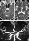
A 60-yr-old woman had suffered from repeated transient right hemiparesis with sensory hypesthesia and mild dysarthria in September 2002. She had a typical pattern characterized by abrupt onset, transient (less than 1 min) duration and spontaneous, complete recovery; this suggested the presence of transient ischemic attack (TIA). The brain magnetic resonance (MR) imaging showed small lacunar infarctions on both thalami and brain stem. The MR angiography showed no definite stenosis or occlusion on any of the large intra- and extra-cranial vessels (Fig. 1). Brain single photon emission computed tomography with an acetazolamide challenge trial showed symmetric perfusion in both cerebral hemispheres. For the diagnosis of TIA, treatment with 75 mg clopidogrel (Palvix™; Sanofi-Aventis, Paris, France) daily maintenance therapy was started. The patient's compliance was good and during the follow-up period, there were no more TIAs, and no side effects were encountered.
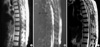
In July 2003, at early dawn, the patient experienced sudden severe back pain, which radiated to her chest. She changed sleeping position but the intensity of pain was not alleviated. In addition, there was subjective weakness in both legs. That morning, the patient was nearly paraplegic; the patient then went to the emergency room. On arrival, her vital signs were stable. The physical examination showed pain and diffuse tenderness along the midthoracic area. Laboratory findings included a hemoglobin value of 11.0 g/dL, white blood cell count of 11,010/µL and a platelet count of 324,000/µL. Bleeding time and coagulation time were 2 min and 5 min, respectively. Neurologic examination revealed paraplegia and severe hypesthesia below the D10 dermatome. Anal tone was not traced, and perianal sensation was not preserved. MR imaging revealed a large elliptical iso- to hypersignal mass occupying the posterior epidural space from T6 to T9 area on T1-weighted images, which changed to CSF-like hypersignal on T2-weighted images. The spinal cord was compressed and displaced anteriorly (Fig. 2). The patient was diagnosed with SSEH.
Nine-hours after paraplegic deterioration, she was underwent emergent laminectomy from T6 to T9; a thick epidural hematoma was evacuated completely. No evidence of vascular malformation was noted at the microscope field as well as at the tissue (hematoma) pathologic examination. Postoperatively the patient improved gradually. Three months postoperatively, her muscle strength was recovered completely. We discontinued the antiplatelet medication and closely followed her for TIA symptoms. During the 1 yr of follow-up, there were no TIA symptoms with the use of gingko extract medication.
DISCUSSION
Patients who have had a TIA or non-disabling ischemic stroke have an annual risk of important vascular events (death from all vascular causes, nonfatal stroke, or nonfatal myocardial infarction) ranging between 4% and 16% from reported clinical trials (7). In the most recent meta-analysis, the Antithrombotic Trialists' Collaboration group reported a 22% reduction of risk, for serious vascular events, in patients receiving antiplatelet therapy (7). Clopidogrel and its precursor ticlopidine are thienopyridines that act via the adenosine triphosphate (ADP) receptors (the P2Y12 receptors) on platelets. The P2Y12 receptors, which are associated with amplification of platelet aggregation and secretion, are irreversibly bound by clopidogrel and inactivated for the life of a platelet (8). Clopidogrel may have some benefit over aspirin in preventing myocardial infarction, ischemic stroke, or vascular death in patients with symptomatic atherosclerosis; its reported major bleeding rate of 1.4% is acceptably low (9).
For patients with spontaneous SSEH, the "spontaneous" refers to "without direct trauma", therefore the pathogenesis may be multifactorial; factors that might contribute to its development include hypertension, ingestion of anticoagulants, straining, sneezing, lifting, and some spinal vascular anomalies (1, 10). Among these potential causative factors, anticoagulant medication may be a predictable one; it has been reported that 25-70% of patients with SSEH have a history of anticoagulant treatment (1). Rarely, SSEH can occur after thrombolytic therapy (11). Antiplatelet agents are frequently used and their hemorrhagic complications occur usually at skin or gastrointestinal sites. Compared to anticoagulants, only four cases of aspirin- and one case of clopidogrel-induced SSEH have been reported (2-6). There is one case of spinal anesthesia-induced spinal epidural hematoma in a patient treated with clopidogrel and enoxaparin (12). We cannot completely rule out the possibility of a coincidence of SSEH and the medical history of taking clopidogrel; however, considering its mechanism of action, the probability of a causal association would seem to be much higher.
The clinical presentation of SSEH has characteristic features of a sudden onset of back or neck pain that radiates to corresponding dermatomes, which is followed by signs of nerve root or spinal cord compression (13, 14). In case of suspicious SSEH, the medical history should address the use of anticoagulants, antiplatelets, or any possible bleeding-inducing agents (15). Compared with the prior use of myelography or CT, MR imaging can diagnose SSEH rapidly and correctly. Therefore, if there is suspicion of a SSEH, an urgent MR imaging should be performed (1). There are a few reports on the spontaneous resolution of SSEH; however, conservative management should be adopted only in patients that have rapid neurologic improvement (4, 13, 16, 17). Close correlation between rapid decompression and postoperative functional outcome make SSEH a critical neurosurgical emergency. According to Groen et al., the operative outcome is totally dependent upon the level of the preoperative neurologic deficit and the operative interval; they reviewed 330 reported cases and concluded that a preoperative incomplete sensory motor deficit correlated highly with a favorable outcome. In addition, recovery was significantly better when decompression was performed in less than 36 hr, in patients with complete sensory motor loss, and in less than 48 hr in patients with incomplete loss (18). Other reports recommended that complete and incomplete cord compression should be relieved within 12 and 48 hr, respectively (1, 14).
In conclusion, with the increasing use of antiplatelet agents, physicians should be alert to this rare but serious complication.
 XML Download