

 PDF
PDF ePub
ePub Citation
 Print
Print


INTRODUCTION
K+ channels play an important role in regulating the membrane potential of smooth muscles and their excitability. Generally, outward K+ current oppose membrane excitability via K+ channels. In a variety of cells including gastrointestinal (GI) tract, several types of K+ channels which are activated by diverse intracellular factors, such as Ca2+ and ATP have been reported (1-7). The best known among them is Ca2+-activated K+ channel (K(Ca) channel) whose gating is regulated by concentration of intracellular free Ca2+ ([Ca2+]i) (3-6). K(Ca) channel participates principally in the rapid repolarization of Ca2+-dependent action potentials (8). Voltage-dependent and Ca2+-independent K+ channels (K(V) channel) and ATP-sensitive K+ channel (KATP channel) in smooth muscles are also known well (1, 7, 9). In GI tract, basal activation of KATP channel has been shown to contribute to control of resting membrane potential (RMP) (9). Recently, pH-sensitive two-pore K+ channels of the TASK family have also been reported in murine GI tract (10). Therefore, it seems that more types of K+ channels still exist in GI smooth muscles. In fact, there is another class of K+ channel not reported in GI smooth muscle cells. This is known as K(Na) channel, which is activated by intracellular Na+ ([Na+]i). It was reported in guinea pig ventricular myocytes (11).
Since its isolation from cardiac myocytes, the gating of K(Na) channel has been known to be activated by intracellular Na+, but not by Ca2+ and ATP or other nucleotides (12). Depending on cell types and recording conditions, reported unitary conductance of K(Na) channel ranges from 105 to 200 pS and half-maximal activation by Na+ occurs between 7 and 80 mM (13). K(Na) channel of the heart was suggested to be activated under pathophysiological conditions such as during failure of the Na+-K+ pump (11). Also K(Na) channel has been proposed to be activated by a single action potential and could be involved in setting RMP in neuronal cell (14-16). To date, however, the existence of K(Na) channel in smooth muscles has not been reported yet. In GI tract, spontaneous contraction and slow wave are dependent on extracellular Na+ concentration: In the duodenum and ileum, low Na+ decreased amplitude, frequency, and rate of rise of the upstroke potentials (17, 18). Acetylcholine (ACh)-induced inward current (IACh) in GI smooth muscles is known as Na+ permeable channel (19). In addition, Na+ current has been reported in human jejunal circular smooth muscle (20). Therefore, it is highly likely that pathways for Na+ influx apparently exist in GI smooth muscles. It is known that intracellular Na+ concentration in resting state reaches less than 10 mM in cardiac and smooth muscle cells (21, 22), and [Na+]i in smooth muscles is increased or expected to increase by the inhibition of Na+-K+ pump (18, 21). These earlier observations suggest a possibility that increased level of intracellular Na+ might play an important role in GI smooth muscle. And one of the possibilities is K(Na) channel. Therefore, this study was designed to prove the existence of IK(Na) in guinea pig gastric myocytes.
MATERIALS AND METHODS
Preparation of cells
Guinea pigs of both gender, weighing 300-350 g, were exsanguinated after stunning. All experiments were performed in accordance with the guidelines for the animal care and use approved by the Chungbuk National University. The antral portion of stomach was cut, and the mucosal layer was separated from the muscle layers in Ca2+-free physiological salt solution (Ca2+-free PSS). The circular muscle layer was dissected from the longitudinal layer using fine scissors and made into small segments (2×3 mm). These segments were incubated in Ca2+-free PSS for 30 min at 4℃. Then, they were incubated for 15-25 min at 35℃ in the digestion medium containing 0.1% collagenase (Wako Pure Chemicals, Osaka, Japan), 0.05% dithiothreitol, 0.1% trypsin inhibitor and 0.2% bovine serum albumin. Following incubation in the enzyme solution, the supernatant was discarded. The softened muscle segments were transferred into Ca2+-free PSS medium, and tissues were washed repeatedly (4-5 times) with Ca2+-free PSS medium. Tissues were then gently agitated with a wide-bore glass pipette to prepare a cell suspension. Isolated cells were kept at 4℃ in Ca2+-free PSS medium until use. Experiments were performed at room temperature within 6 hr of harvest of cells.
Voltage-clamp patch experiments
Isolated cells were transferred to a small chamber on the stage of an inverted microscope (IX-71, Olympus, Tokyo, Japan). The chamber was perfused with PSS (2-3 mL/min). Glass pipettes with a resistance of 2-5 MΩ were used to make a giga seal of 5-10 GΩ, by using standard patch clamp techniques (23). Membrane currents were amplified with an axopatch-1C or 200B patch-clamp amplifier (Axon Instruments, California, U.S.A.), and a data were digitized with Digidata 1,220 or Digidata 1,322 and stored directly and digitized on-line using pClamp software (version 5.5.1 or 9.2). Data were displayed on a digital oscilloscope, pen recorder (Gould, Cleveland, OH, U.S.A.) and a computer monitor, and data were analyzed using pClamp 6.0 (pClamp 9.2) and Origin software 6.1.
Solution and drugs
Ca2+-PSS, containing (in mM) NaCl 140, KCl 5, CaCl2 2, MgCl2 1, glucose 5, and HEPES (N-[2-hydroxyethyl] piperazine-N'-[2-ethanesulphonic acid]) 10, and pH was adjusted to 7.4 with NaOH. CaCl2 was simply omitted in the Ca2+-free PSS. Equimolar concentrations of external Na ([Na+]o) were replaced by K+ for various extracellular K+ ([K+]o). In some experiments, [Na+]o and [K+]o were replaced by 145 mM N-methyl-D-glucamate chloride (NMDG) or CsCl2. Pipette solution, containing (mM) KCl 145, GTP (Tris form) 0.1, EGTA (ethylene glycol-bis (2-aminoethyl ether- N,N,N',N'-tetraacetic acid) 10, creatine phosphate (CrP, Tris form) 2.5, HEPES 10, ATP (Tris form) 4, MgCl2 5, and pH was adjusted to 7.3 with TRIZMA. Equimolar concentration of K+ in K+-rich pipette solution was replaced by Li+ or Na+ for making Li+- and Na+-rich pipette solution, respectively. For low Cl- pipette solution, containing Na-gluconate 100, KCl 13, Na2GTP 0.1, Na2ATP 2.5, Na2CrP 2.5, EGTA 10, HEPES 10, Mg2ATP 1.5, MgCl2 3.5, K -gluconate 22, pH was adjusted to 7.3 with TRIZMA. R56865 (N-[1-(4-(4-fluorophenoxy) butyl)-4-piperidinyl]-N-methyl-2-benzothiazolamine was a kind gift from Janssen Research Foundation, Beerse, Belgium). All drugs used in this study were purchased from Sigma (St. Louis, MO, U.S.A.).
RESULTS
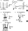
Membrane potential was held at -60 mV, and hyperpolarizing-ramp pulse, ranging from 80 to -120 mV (-0.05 V/sec), was applied to cells with 15 sec interval. After the whole-cell configuration was established, inward current was produced by increasing the external K+ from 4.5 mM to 140 mM. The amplitude of the steady-state inward current was -47±13.1 pA under K+ rich pipette solution (n=4, Fig. 1Aa). However, significantly large amplitude of the steady-state inward current was produced by increasing [K+]o (60 mM) under 110 mM of intracellular Na+ concentration ([Na+]i) (-1,052±145.7 pA, n=5, Fig. 1Ab). Right panel of Fig. 1Ab shows the current/voltage (I/V) relationship of Na+-induced inward current by increasing [K+]o (60 mM) under 110 mM [Na+]i ([K+]i=35 mM). Reversal potential (Erev) of this current in I/V relationship was 12.5 mV and it was close to expected K+ equilibrium potentials (13.6 mV). Fig. 2A shows I/V relationship of Na+-induced inward current by increasing [K+]o (60 and 140 mM) under 80 mM [Na+]i. Erev of each current in I/V relationship was 1 and 19 mV, respectively (Fig. 2A). Each potential was shifted to rightwards, following expected potentials by increasing [K+]o (60 and 140 mM). These inward currents which was activated by increasing [K+]o was blocked by total replacement of external solution with [NMDG+]o and [Cs+]o, respectively (Fig. 1B).
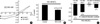
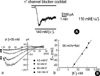
Effects of K+ channel blockers, such as Ba2+, glibenclamide, tetraetylammonium (TEA), 4-aminopyridine (4-AP), apamine, and quinidine, on the steady-state of Na+-induced current were studied. Either of Ba2+ (100 µM), glibenclamide (10 µM), 4-AP (10 mM), or quinidine (3 µM) did not block Na+-induced inward current (p>0.05; data not shown). However, TEA (5 mM) decreased the steady-state current of Na+-induced current to 68±2.7% of the control (n=5, p<0.05, Fig. 2B). Since K(Ca) and KATP channels are present in GI tract, Na+-induced inward current was studied in the presence of diverse K+ channel blockers to eliminate the possibility of contamination with other types of K+ channels. In the presence of K+ channel blocker cocktail (mixture of K+ channel blockers using Ba2+ [100 µM], glibenclamide [10 µM], TEA [10 mM], 4-AP [10 mM], apamine [300 nM], and quinidine [3 µM]; pH was adjusted to 7.4 before use), Na+-dependent current was still recorded. The amplitude of inward current with 140 mM [K+]o was -1,063±182.1 pA (Fig. 2C, 3A). However, small steady-state inward current (-60±38.1 pA) was shown under Li+-rich pipette solution (n=4, p<0.05; Fig. 2C). It has recently been reported that certain types of KNa channels (slick and slack channels) have a Cl- dependence (24). To evaluate the Cl- dependence of Na+-induced inward current in guinea pig gastric myocytes, amplitude of the Na+-induced inward current was compared using 20 mM Cl- pipette solution. The amplitude of Na+-induced inward current under 20 mM Cl- and 110 mM [Na+]i in the presence of K- channel blocker cocktail was -471±86.0 (n=5, Fig. 2C). To evaluate the ionic selectivity of inward current obtained in the presence of K+ channel blocker cocktail, Erev was also determined from the I/V relationship and plotted against logarithmic scale of [K+]o (Fig. 3Bb). When the line-fitting was applied to the data points, a slope of recorded data under K+ channel blocker cocktail and low Cl- pipette solution was obtained. In 3 tested cells, the slope was 52±2.5 mV per 10-fold change in [K+]o. These results suggest that Na+-induced inward current in guinea pig gastric myocytes is selective to K+ ions.
Finally, to confirm that Na+-induced inward current is IK(Na), we studied the effect of R-56865 which is known to block IK(Na). As shown in Fig. 4A, R-56865 inhibited Na+-induced inward K+ current in a reversible manner. Ten and twenty µM R-56865 inhibited Na+-induced inward current to 11±5.7 and 4±2.9% of the control (n=3 and 5), respectively (p<0.05, Fig. 4B) in the presence of K+ channel blockers and low Cl- pipette solution.
DISCUSSION
Intracellular Ca2+ is regarded as one of main regulatory factor in many biological responses including activation of ion channels (6). In contrast to Ca2+, Na+ has not generally been considered as an important intracellular messenger to say nothing of muscular cells. However, since K(Na) channel was first identified in guinea pig cardiac myocytes, its existence and importance of [Na+]i have also been reported in various neuronal cells (11, 12, 15, 16, 25). These facts naturally raise a question of whether K(Na) channel exists in GI smooth muscle. As can be seen in Fig. 1Aa, increasing [K+]o (140 mM) failed to produce inward current without Na+ in pipette solution. This is also true when Na+ in pipette solution was replaced with Li+ in the presence of K+ channel blocker cocktail (Fig. 2C). However, addition of Na+ into pipette solution produced inward current by stimulation of increasing [K+]o. As can be seen in Fig. 1Ab (110 mM [Na+]i) and 2A (80 mM [K+]o), increasing [K+]o produced larger inward currents, and the Erev was moved to rightward direction following the values expected. However, this current was not affected by pretreatment with ouabain (10 µM) known to blocker of Na+-K+ pump (data not shown).
Even though K(Na) channel has been reported to exist in the heart and neurons, its electrophysiological and molecular properties are poorly understood compared with other K+ channels. Furthermore, characteristics of K(Na) channel, have not yet been reported in any smooth muscle including GI tract. To date, two representative molecular identities known as KNa channel subunits have been reported: Slack and Slick gene families (24, 26). In both type of KNa channels, TEA (20 mM) was reported to inhibit KNa channels significantly (24). As seen in Fig. 2B, TEA (5 mM) significantly inhibited Na+-induced current in guinea pig gastric myocytes. Since K(Ca) and KATP channels are present in GI tract, we tried also to exclude activation of K(Ca) and KATP channels by high Ca2+ buffering capacity, using 10 mM EGTA and 4 mM ATP, at holding potential of -60 mV. In addition, to prevent possible activation of other types of K+ channels, we studied [Na+]-induced current in the presence of K+ channel blocker cocktail which contained Ba2+ (100 µM), glibenclamide (10 µM), TEA (10 mM), 4-AP (10 mM), apamine (300 nM), and quinidine (3 µM) (Fig. 2C, 3A). In the presence of K+ channel blocker cocktail, Na+-induced inward current was activated, although the amplitude of the steady state current was reduced. As shown in Fig. 2C, IK(Na) was further reduced under low Cl- in the presence of K+ channel blocker cocktail. It has recently been reported that certain types of KNa channels (slick and slack channels) are Cl- dependent and slick is highly sensitive to Cl- (27). As shown in Fig. 2C, 3Ba and 4A, the amplitude of Na+-induced current in the presence of K+ channel blocker cocktail was significantly reduced by low Cl- pipette solution. However, as shown in Fig. 1B, Na+-induced inward current was blocked by 140 mM [NMDG+]o and [Cs+]o. Therefore, it seemed that IK(Na) in guinea pig gastric myocytes might also be Cl--dependent for its activation.
Ionic selectivity of Na+-induced current was studied under low Cl- pipette solution in the presence of K+ channel blocker cocktail to eliminate all possible contaminations. As shown in Fig. 3Bb, the slope of reversal potential obtained versus [K+]o in the presence of K+ channel blocker cocktail by linear-fitting was 52±2.5 mV per 10-fold change in [K+]o (28). Finally, Fig. 4 shows reversible inhibitory effect of R-56865 (10 and 20 µM) on Na+-induced inward current. These results are very similar to the results observed in cardiac cells (29, 30). All the above findings suggest that Na+-induced inward current recorded in our experiment is IK(Na) found in guinea pig gastric myocytes. For activation of K(Na) channel of cardiac myocytes in single channel level, it required at least 10-20 mM [Na+]i (11, 12, 16). A recent report also suggests that the half-maximal activation of K(Na) channel by Na+ occurs between 7 and 80 mM, depending on cell types and recording conditions in single channel levels (13). Therefore, single channel study appears to be required in the future to determine the threshold of [Na+]i for activation of IK(Na) in guinea pig gastric myocytes.
In cardiac myocytes, it was reported that normal level of [Na+]i is less than 10 mM (22). Therefore, it is unlikely that K(Na) channel would ever be activated under normal conditions. Instead, a considerable increase of [Na+]i is possible in case of a prolonged failure of Na+ pumping after the onset of acute ischemia or during anoxia. Increase of [Na+]i would cause reversal of Na+-Ca2+ exchange due to a reduced driving force for Na+ ions (11, 31, 32). However, the activation of IK(Na) following intracellular Na+ accumulation may set membrane potential to more negative ranges so that Na+-Ca2+ exchanger may maintain the driving force for Na+ influx, thereby improving Ca2+ transport (33). In the case of neurons, [Na+]i in resting state also ranges between ~4 and 15 mM (27), however, [Na+]i in neurons can also reach as high as ~100 mM by stimuli in dendrites (27, 34). Therefore, it has earlier been suggested that the activation of IK(Na) could be involved in setting RMP and generating the spindle waves of neurons (15, 16, 35). As known well, there are several Na+ influx pathways in GI tract such as Na+ channel, IACh and Na+-K+ pump (18-21). Therefore, IK(Na) may play an important physiological role in the regulation of membrane potential, hence the motility of GI tract. Since there has been no report until now on the identification of K(Na) channel in either GI tract or whole smooth muscle research field, the present study is the first report to prove the existence of IK(Na) in guinea pig gastric myocytes.

 XML Download