

 PDF
PDF ePub
ePub Citation
 Print
Print


INTRODUCTION
The prolonged presence of retrophayngeal abscess can cause cervical spinal epidural abscesses, which require urgent treatment Epidural extension of a retropharyngeal abscess is an extremely serious but very rare condition (1). Retropharyngeal abscess occurs mostly in the thoracic and the lumbar areas (2). The main pathogenesis of these conditions is hematogenous seeding to the epidural space from a distant source of infection. However, penetrating trauma especially caused by fish bones, can also be the cause (3). The causative organisms of spinal epidural abscesses are the common species of Staphylococcus or Streptococcus.
Eikenella Corrodens is part of the normal flora of the mouth and the upper respiratory tract. Infections by this organism occuring in association with bite wounds, fist-fight injuries or dental and periodontal inflammations are well known.
We report a case of E. corrodens cervical spinal epidural abscess induced by fish bone.
CASE REPORT
A 72-yr-old man with hypertension, diabetes, and chronic obstructive pulmonary disease presented with a history of right side motor weakness and dyspnea for 3 days. He stated that he had a fish bone had in his throat for about 2 months. The patient did not seek medical attention and experienced continuous pain in the posterior neck area and both shoulders. On admission, he was alert and cooperative. Physical examination revealed moderate nuchal rigidity and tenderness along the cervical spine with marked restriction of neck motion. Blood pressure was 145/86 mmHg, pulse rate was 97 beats/min, and oral temperature was 37.8℃. On neurological examination, Lhermitte's sign was positive, and anisocoric pupil size (2/3 mm) and anhydrosis on the right side of his face were detected. Concerning the motor and sensory systems, right hemiparesis and left hypesthesia were observed, while deep tendon reflexes were normal. Also, pathologic reflexes were absent and the patient did not have voiding difficulty. The patient was examined for medullar and cervical cord lesions.
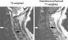
Laboratory findings showed a leukocytosis of 12,000/µL, an ESR of 38 m/hr, and CRP of 2.16 mg/dL. Simple radiography showed retrolisthesis of the 3rd cervical spine, cervical spondylosis, and scoliosis. Cervical MRI showed continuous signal changes from medulla to cervical cord and a retropharyngeal abscess with osteomyelitis in the C3 and C4 vertebral bodies (Fig. 1). Gastro fiberscopy performed before surgery showed neither signs of a penetrating fish bone nor related injuries.
On the 2nd hospital day, the patient started to show respiratory distress. Emergent surgery was performed with corpectomy of C3-C4 vertebral body, and then epidural space abscess was drained. Finally, anterior cervical interbody fusion using autologous bone graft obtained from the patient's iliac crest was performed. A skate bone was found at the C3-C4 intervertebral disc space (Fig. 2). Intraoperative cultures, as well as pathologic specimens, were obtained. The sample was inoculated onto 5% sheep blood agar plate as well as onto chocolate blood agar plates. A pure growth of E. corrodens was obtained after 48 hr of incubation at 37℃ in a carbon dioxide-enriched atmosphere.
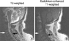
After the surgical debridement, the patient was treated with intravenous ciprofloxacine 400 mg twice a day for 6 weeks. Despite aggressive antibiotic therapy and surgical drainage, the right hemiparesis remained and left paresthesia only slightly improved. After 4 months, cervical MRI showed a decrease of swelling, signal intensity of spinal cord, retropharyngeal abscess and spondylitis compared to the previous cervical MRI (Fig. 3).
The patient was treated for pneumonia during his hospitalization and was discharged on the 371st hospital day. He is now on rehabilitation therapy at an outpatient clinic.
DISCUSSION
The cervical area is the rarest site of spinal epidural abscesses (2, 4), with more common locations being the lower parts of the spine. However, cervical abscesses seem to exert the most devastating effects. More than one third of patients who survive this illness are left with major neurological deficits, mainly paralysis. In our case, right hemiparesis and left hypesthesia remained.
It has been well-documented that infections of the vertebral body are caused by many different mechanisms (5). Most commonly, spinal epidural abscesses occur from hematogenous spread. Direct contact with bacteria from foreign material related to spinal surgery, injection of dye into disc space, epidural steroid injection, spinal or paravertebral anesthesia and lumbar puncture may also lead to infection of the spine. Among these mechanisms, retropharyngeal abscesses usually develop into a pharyngeal infection. Regional trauma from an ingested foreign body is the cause in 59% of patients with a retropharyngeal abscess. Only a small number of retained foreign bodies (such as fish bones, wire, meat bones, and chicken bones) can perforate the upper digestive tract lumen, and an even smaller fraction completely migrates extraluminally (6). Also, the incidence of a retropharyngeal abscess associated with an epidural abscess is very rare (7).
The causative organisms found in epidural abscesses are most commonly Staphylococcus or Streptococcus. E. corrodens has not been reported as a causative organism of a retropharyngeal and spinal epidural abscess. E. corrodens is fastidious, facultative anaerobic, gram-negative bacillus that pits or corrodes the surface of solid culture media. The organism is part of the normal flora, grows slowly, and does not cause hemolysis, hydrolyze starch, form indole, or ferment monosaccarides. Growth of E. corrodens is enhanced by blood agar media, x-factor (hemin), and exposure to an atmosphere with 3-10% carbon dioxide (8). Antecedent trauma and systemic factors such as advanced age, malignant neoplasms, drug addiction, altered host immunity, and recent dental manipulation occasionally predispose a person to infections with E. corrodens.
In this case, there were no dental procedures, dental infections, or any prior bite wounds, nor any other predisposing factors except for diabetes, hypertension, and chronic obstructive pulmonary disease.
Cervical spinal epidural abscesses can cause considerable neurological disability. The progress of this infection through its stages of spinal ache, root pain, weakness, and eventual paralysis has been well-described. However, early diagnosis remains difficult, since a large number of patients are initially released from emergency rooms without treatment (9). A detailed history about past medical problems and relevant events is critical for the early diagnosis and timely management.

 XML Download