

 PDF
PDF ePub
ePub Citation
 Print
Print


INTRODUCTION
Racemic bupivacaine (RBUP) can cause life-threatening cardiac complications, particularly when given intravascularly by accident. Levobupivacaine (LBUP) was developed as a long acting local anesthetic with a potentially reduced toxicity compared with RBUP. LBUP is considered to be safer than RBUP because it has a higher cardiotoxicity threshold and lower risk of CVC (1). However, by the direct intracoronary infusions in conscious sheep, both RBUP and LBUP caused similar fatal cardiotoxicity (2). The incidence of electrical stimulation-induced ventricular tachycardia and fibrillation with LBUP was similar to that with RBUP (3). A recent report showed that a relatively small amount of LBUP (approximately 1.6 mg/kg) could lead to CVC when infused into the systemic circulation in an anesthetized human (4). Furthermore, regarding the successful resuscitation with a conventional treatment, the LBUP-induced CVC failed to show significant advantage over the RBUP-induced CVC (5, 6).
The LBUP-induced CVC may be different in the degree of not only cardiac depression but also vascular reactivity from the RBUP-induced CVC, as CVC is caused by cardiac depression in association with vascular collapse. In spite of less cardiac depression, the vascular reactivity may be more compromised by LBUP, thereby the resuscitation from the CVC caused by LBUP may be as difficult as that by RBUP in some studies (5, 6).
The resuscitation from RBUP-induced CVC with conventional treatment is known to be difficult, so many modalities have been applied for better results. Cho et al. (7) and Kim et al. (8) proposed the feasibility of insulin for the resuscitation from the RBUP-induced CVC. Considering the channel effect (9) and cardiac supportive action of insulin (10), it can be assumed that insulin may work on the LBUP-induced CVC.
We hypothesized that insulin may be effective in resuscitating the CVC by either bupivacaine, and that there may be a different pattern of recovery between the two bupivacaines. The possible difference in the cardiac depression and vasoactivity may be augmented when insulin, which has vasodilatory activity in addition to positive inotropic action (11), is used as a resuscitative agent. This study was performed to compare the insulin-mediated recovery from the LBUP-induced CVC with that from the RBUP-induced CVC, and to assess the feasibility of insulin resuscitation for the LBUP-induced CVC.
MATERIALS AND METHODS
The study was approved by the committee of animal care and use of our institute. Enrolled were fourteen male mongrel dogs weighing between 19 and 32 kg. They were randomly assigned to two groups (n=7 for each); the RBUP and LBUP group. Commercial forms of RBUP (Bupivacaine HCL Inj., Myungmun Pharmacy, Korea) and LBUP (Chirocaine®, Abbott Laboratories Ltd., Ireland) were used for the induction of CVC in each group.
Anesthesia was induced using thiopental sodium (10 mg/kg) and maintained by continuous sodium pentobarbital infusion (5 mg/kg/hr) and vecuronium injection (0.2 mg/kg initially and 0.02 mg/kg at 30-min intervals). The dogs were tracheally intubated and ventilated with 100% O2 maintaining normocarbia. Volume requirement during NPO time was calculated and administered over the first half hour. Additional volume support with normal saline was initiated from 2 to 8 mL/kg in response to signs of hypovolemia during the stabilization period. A fluid infusion of 2 mL/kg/hr was maintained to account for insensible losses throughout experiments. Body temperature was kept at 37-38℃ and sodium bicarbonate was administered (2-4 mEq/kg/hr) to control the acid-base balance (pH 7.35-7.45).
Two antecubital venous catheters were used to infuse drugs and normal saline. Both femoral arterial catheters were cannulated to monitor arterial pressure continuously and to collect blood samples. A Swan-Ganz catheter (Opticath®, P7110-EH, Abbott, U.S.A.) was introduced via the external jugular vein to measure central venous pressure (CVP) and cardiac output (CO).
After a 30-min stabilization period, RBUP or LBUP infusion was started at a rate of 0.5 mg/kg/min (0.5% mixture, both drugs) until the mean arterial pressure (MAP) reached 40 mmHg, which was defined as the time of CVC as we have found that no dogs in two groups could survive spontaneously at that point in our preliminary study using three dogs in each group. At CVC, RBUP or LBUP infusion was stopped and 2 U/kg of regular insulin (Novolin® R, Novo Nordisk, Denmark) was injected via a central venous catheter followed by the infusion of D50W (2 mL/kg for 30 min) and potassium (1-2 mM/kg/hr).
The criterion for successful resuscitation was as follows; 90% of initial MAP should be achieved by 30 min following cardiovascular collapse and be maintained at that level for a minimum of 30 min.
All data were collected until 60 min after CVC. MAP and heart rate were continuously monitored and recorded at 1-min intervals using a HP Component Monitoring System™ (Hewlett-Packard Model 54S, Andover, MA). CO was recorded as a mean of two consecutive measurements every 5-min throughout the experiment using the thermodilution technique. Systemic vascular resistance (SVR) was calculated using the standard formula (SVR=80×[MAP-CVP]/CO). ECG analysis was performed every 5-min using a resting ECG analysis system (MAC8®, Marquette, U.S.A.). Arterial blood samples were collected to determine immediate blood gas, electrolytes, and glucose levels every 10-min through the experiment. Blood samples were centrifuged at 2,500 rpm for 15 min to determine total plasma drug concentrations by high performance liquid chromatography using an ultraviolet detector operation at a wavelength of 204 nm with a limit of sensitivity of 4 ng/mL.
Data were expressed as mean±SD. The independent samples T-test and the Mann-Whitney U-test were used for analysis of between-group differences. Recovery phase values were compared to the baseline values or to CVC values using the Wilcoxon signed rank test and the Sign test with Dunnette or Tukey post hoc testing. Time-dependent variables were analyzed by repeated measures of ANOVA. All statistical analyses were performed using SPSS software (Version 10.09), and p<0.05 was considered significant.
RESULTS
There were no differences in weight, time to CVC, drug dose required for CVC, and total plasma drug concentration at CVC between the two groups (Table 1).
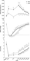
CVP increased continuously until CVC but was not different between two groups (5±2 mmHg at baseline and 11±2 mmHg at CVC). The infusion of bupivacaines decreased CO continuously with the lowest value at CVC in both groups. CO was higher in the LBUP group than in the RBUP group at CVC (p<0.05). Calculated SVRs increased at CVC in both groups but the SVR was significantly lower in the LBUP group than in the RBUP group (p<0.05) (Fig. 1).
After insulin injection, all dogs in both groups were successfully resuscitated with restored baseline blood pressures, but time spent under 40 mmHg during resuscitation was significantly longer in the LBUP group (p<0.05). MAP declined further in both groups for minutes and then restored 40 mmHg at 3.4±0.5 min after CVC in the RBUP group and at 6.1±1.2 min in the LBUP group (Table 1). The CO declined in the RBUP group in excess of that observed in the LBUP group, and the CO remained lower in the RBUP group at each measured time interval. The SVR was higher in the RBUP group from CVC for a period of 20 min, and then both groups recovered the baseline SVR 40 min after insulin injection (Fig. 1).
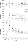
In addition to arrhythmias, which manifested as sinus arrhythmia or 1st degree AV block in the LBUP group and premature ventricular complex or a supraventricular beat in the RBUP group immediately prior to CVC, infusion of bupivacaines produced prolongations of PR and QRS intervals, which were less in the LBUP group. The restoration of ECG parametric values during recovery was only seen in the QRS interval in both groups (Fig. 2).
Blood sample analyses, which included acid-base balance (pH 7.35-7.41), plasma potassium (3.7-4.4 mM), and blood glucose (99-108 mg/dL), were within physiologic ranges throughout the experiment, and between-group differences were not observed. Total plasma bupivacaine concentrations showed no difference between the two groups throughout the experiment. These concentrations reduced abruptly after insulin injection (at 5 min after insulin injection, 12.2±1.6 mcg/mL in the LBUP group, and 7.9±1.2 mcg/mL in the RBUP group) and continued to decrease until the end of the experiment.
DISCUSSION
Management of local anesthetic-induced CVC is aimed at correcting contractile dysfunction of the heart and arrhythmia. When some confounding factors such as hypoxia, hypercarbia, acidemia, and hyperkalemia were present, there were increased incidences of arrhythmia with RBUP (12, 13). However, we could not determine the effect of arrhythmia on CVC in this study because arrhythmia and ECG parametric changes induced by two bupivacaines did not progress towards hemodynamic instability when eliminating confounding factors mentioned above. Liu et al. noticed that 'pump failure' was the cause of death in the RBUP-treated anesthetized dogs (14). We speculate that the primary cause of CVCs induced by two bupivacaines may be hypotension from diminished contractilities rather than arrhythmias in anesthetized dogs.
Most studies evaluating resuscitative modalities were performed using RBUP, whereas only a few studies used LBUP because LBUP has been considered safer than RBUP. There is indirect evidence from binding kinetics to suggest that recovery from cardiotoxicity is faster with LBUP than with RBUP in the guinea-pig heart (15). Moreover, following the intravenous infusion of LBUP or RBUP in conscious sheeps, arrhythmias due to LBUP returned to sinus rhythm more readily than those due to RBUP (16). However, the parametric changes of ECG were not outstandingly different from each other, and the CVC induced by LBUP was as profound as that by RBUP in our study. The potential advantage of LBUP over RBUP was not observed in a recent study by Groban et al. in anesthetized dogs (5). After infusion of local anesthetics to the point of CVC, hypotension and arrhythmias were treated with epinephrine, open-chest massage, and advanced cardiac life support protocol, respectively. They concluded that, after RBUP or LBUP infusion, resuscitation is not always successful and co-administration of epinephrine may lead to severe ventricular arrhythmias. The CVC by LBUP seems to require an intensive treatment such as like the CVC caused by RBUP.
Animal studies have shown that resuscitation using sympathomimetics, particularly norepinephrine and epinephrine, improves outcome in RBUP-induced asystole (17). However, epinephrine may exacerbate arrhythmias associated with local anesthetic overdose without improving CO (18). Phosphodiesterase inhibitors having both positive inotropic and vasodilatory actions are expected to improve CO. However, they do not necessarily support blood pressure, and are associated with a significant incidence of ventricular arrhythmias (19).
Cho et al. reported that, after the infusion of RBUP in dogs, treatment with a combination of insulin and glucose facilitated recovery of blood pressure, cardiac output, and ECG parameters (7). They supposed that intracellular K+ transport by insulin counteracts RBUP inhibition of transient outward current improving rates of myocardial repolarization. They also proposed that insulin might have an effect on calcium homeostasis and sodium channel dynamics, and that catecholamine release might account for the benefit of insulin resuscitation. Weinberg and VadeBoncouer proposed the energy-related mechanism of insulin during resuscitation stating that insulin's cardiac supportive effect is due to promoted use of fatty acid as a metabolic source by the ischemic heart (20). We postulated that insulin might show such a favorable result in resuscitation from the LBUP-induced CVC as in the RBUP-induced CVC, if the same mechanism worked when the heart remained in a state of extreme depression.
Insulin seems to be effective for the resuscitation from the LBUP-induced CVC but with substantial delays. In dogs, increases in plasma insulin levels to high physiological concentrations did not increase blood pressure because increases in cardiac output were offset by decreases in vascular resistance (21). Similarly, in humans, short-term insulin infusion failed to increase arterial pressure possibly by the same mechanism (22). In this study, the vasodilatory effect was also seen following administration of insulin, but a discrepancy existed between two groups; in spite of higher cardiac output, the restoration of MAP in the LBUP group was more protracted than that in the RBUP group. We infer that LBUP is less cardiac depressive than RBUP, but vascular reactivity to compensate the cardiac depression may be more compromised in the presence of LBUP. The unfavorable results from previous reports on the resuscitation from the LBUP-induced CVC might have been due to the fact that LBUP has a relatively more vasodilatory property than RBUP and the vasoactivity plays a role in the CVC induced by LBUP. In this study, the discrepancy in immediate restoration of blood pressure seems to be due to the difference in vascular reactivity between the two bupivacaines, which was combined by the systemic vasodilatory effect of a large bolus of insulin.
Shaffner et al. demonstrated the importance of early institution of cardiopulmonary resuscitation because a delay beyond 6 min markedly impairs the ability to generate viable levels of cerebral blood flow in dogs (23). Furthermore, even at cerebral perfusion pressure of 35 mmHg after 6 mins of CVC, cerebral metabolic recovery is incomplete and inadequate to restore full reperfusion. The perfusion pressure and time, less than 40 mmHg for 6 min during the early resuscitation period in the LBUP group, is comparable with their results, emphasizing the risk of recovery lag.
Insulin may be a good choice in managing the RBUP-induced CVC because the severely depressed heart generally responds negatively to increased afterload and insulin may facilitate the recovery by lowering the afterload. However, based on our findings, the management strategy of the LBUP-induced CVC may differ from that of the RBUP-induced CVC. Insulin can be effectively used for treating the LBUP-induced CVC, but insulin or any inotropes with a vasodilatory activity may need adjunctive therapy to abolish the hypotensive recovery lag during the immediate resuscitation period because of lowered SVR.
In conclusion, a single injection of insulin can resuscitate CVCs induced by LBUP or RBUP in anesthetized dogs, but the innate vasodilatory property of insulin in combination with the less increased SVR delays the restoration of blood pressure during the immediate resuscitation period in the LBUP-induced CVC.

 XML Download