

 PDF
PDF ePub
ePub Citation
 Print
Print


INTRODUCTION
Food allergy is now accepted as a major health problem worldwide, especially in westernized nations. Food allergy is an adverse immunologic reaction that might be due to IgE- or non-IgE-mediated immune mechanisms and is known to cause gastrointestinal allergy, atopic dermatitis, and respiratory allergic diseases. Although intact food allergens routinely penetrate into the gastrointestinal tract, clinical symptoms of allergy rarely develop because tolerance to food antigens is established in most individuals. However, there are individuals with atopic tendency who become sensitized to several food antigens and ultimately develop allergic symptoms (1, 2).
Buckwheat (BW) (Fagopyrum esculentum) is a major cause of food hypersensitivity in children in Korea and Europe. In 1909, Smith first described a young patient presenting with severe symptoms of asthma, allergic rhinitis, urticaria, and angioedema after ingesting a small quantity of BW (3). House dust mite (HDM) is the most important aeroallergen worldwide (4); 70% of pediatric pulmonologic patients and half of adult patients in Korea are known to have been sensitized to HDM, Dermatophagoides farinae (Der f) and Dermatophagoides pteronyssinus (Der p), based on skin tests (5). Nickel et al. showed that risk factors for sensitization to indoor and/or outdoor allergens at 3 yr were a positive family history and the presence of hen's egg-specific IgE antibodies (≥0.35 kU/L) at the age of 12 months (6). Therefore, sensitization to food allergens early in life is thought to be associated with allergic asthma in children and adults and the development of IgE-mediated hypersensitivity to inhalant allergens (7). However, despite the bulky work investigating allergy, no murine model possessing the characteristics of both food and inhalant allergy has been established.
In the present study, we addressed the influence of BW exposure on IgE-mediated hypersensitivity response to HDM by measuring serum BW- and HDM-specific IgE antibodies. Moreover, the role that T cells play in the regulation of BW and HDM allergy was explored by measuring cytokine production by splenocytes from mice allergic to BW and/or HDM.
MATERIALS AND METHODS
Animals and reagents
Female C3H/HeJ mice, 4 weeks of age, were purchased from SLC Japan (Hamamatsu, Japan) and maintained on regular mouse chow (BW-free chow) under specific pathogen-free conditions. Guidelines for the care and use of the animals were followed (8). Fresh BW and HDM crude extracts were used as antigens. Crude BW was obtained from the Korean Rural Development Administration. Crude HDM was obtained from the Department of Parasitology, Yonsei University College of Medicine. The crude extracts were prepared as described below. CT was purchased from List Biological Laboratories, Inc. (Campbell, CA, U.S.A.) and concanavalin (Con A) from Sigma (St. Louis, MO, U.S.A.). Antibodies for ELISAs were purchased from PharMingen (San Diego, CA, U.S.A.) and anti-DNP IgE from Accurate Scientific Inc. (Westbury, NY, U.S.A.).
BW crude extract preparation
Freshly ground whole BW and crude BW extract were prepared as previously described (9) and used as antigen. Briefly, 50 g of BW flour was defatted with ethyl ether and then extracted in 500 mL of phosphate-buffered saline (PBS, 137 mM NaCl, 1.8 mM KH2PO4, 10 mM Na2HPO4, 27 mM KCl, pH 7.4) for 24 hr at 4℃ under constant stirring. The extract was centrifuged at 10,000 g for 1 hr at 4℃, and the supernatant was dialyzed (the cut-off molecular weight was 3.5 kDa; Spectrum, Houston, TX, U.S.A.) against distilled water for 48 hr. The dialyzed supernatant was lyophilized and stored at -20℃ until use.
HDM crude extract preparation
Freshly ground HDM and crude HDM extract were prepared as previously described (9) and used as antigen. Briefly, 10 g of HDM was defatted with ethyl ether and then extracted in 500 mL of phosphate-buffered saline (137 mM NaCl, 1.8 mM KH2PO4, 10 mM Na2HPO4, 27 mM KCl, pH 7.4) for 72 hr at 4℃ under constant stirring. The extract was centrifuged at 50,000 g for 1 hr at 4℃, and the supernatant was dialyzed (the cutoff molecular weight was 1 kDa; Spectrum, Houston, TX, U.S.A.) against distilled water for 48 hr. The dialyzed supernatant was lyophilized and stored at -20℃ until use.
Sensitization by intragastric administration of BW
Mice were sensitized intragastrically with BW plus CT as an adjuvant on days 0, 1, 2, 7 and 18 (Group 1, n=4) (Fig. 1). Two hours before intragastric sensitization was done, mouse chow was removed. Intragastric feeding was performed by means of a stainless steel blunt feeding needle. To establish the optimum sensitizing dose, mice were given 1 mg (low dose) of BW together with 10 µg/mouse of CT. Preliminary studies revealed that 1 mg/mouse of BW plus 10 µg/mouse of CT is most effective in provoking increases in IgE and IL-4 levels (data not shown). The BW/CT mixtures were administered in PBS at a final volume of 200 µL/mouse. Control mice received CT alone (Group 2, n=4) or were left untreated (Group 3, n=4).
Sensitization by inhaled administration of HDM
Mice in Groups 1 and 2 were sensitized with 200 µg/dose of crude HDM extract plus 400 µg/dose of Al (OH)3 as an adjuvant through intraperitoneal routes (on day 0). Two weeks later (on days 14, 15, 16, and 21), mice in Groups 1 and 2 were given 100 µg intranasal boost of HDM (Fig. 1). Blood samples were taken following exsanguinations on day 35. Fig. 1 illustrates an overview of the study groups.
Measurements of BW- and HDM-specific IgE, IgG1, and IgG2a in sera
Blood was obtained weekly from the tail veins of the mice during the sensitization period and was taken following exsanguinations on day 35. Sera were collected and stored at -20℃. Levels of BW- and HDM-specific IgE were measured by ELISA as previously described (10). For measurements of BW- and HDM-specific IgG1 and IgG2a, Maxisorp Immuno 96-well plates (Nunc, Denmark) were coated with either 2 µg/mL BW or HDM in coating buffer (pH 9.6, Sigma, U.S.A.). After overnight incubation at 4℃, plates were washed 3 times with PBS/0.05% Tween 20 and blocked with 1% BSA-PBS for 2 hr at 37℃ After washing 3 times, serum samples (1:10 dilutions for IgE, 1:500 dilutions for IgG1 and IgG2a) were added to the plates and incubated overnight at 4℃. Plates were then washed, and 100 µL of secondary antibody (antimouse IgE, IgG1, or IgG2a) conjugated with biotin (0.5 µg/mL) were added for an additional 1 hr at room temperature (RT). After washing, avidin peroxidase (PharMingen, San Diego, CA, U.S.A.) was added to wells for 15 min at RT. After 6 washings, reactions were developed with TMB substrate (PharMingen, San Diego, CA, U.S.A.) for 30 min at RT, stopped with 2 N H2SO4, and read at 450 nm. The levels of antigen-specific IgE, IgG1, and IgG2a were calculated by comparison with a reference curve generated by using mouse mAbs (anti-DNP IgE, IgG1, and IgG2a), as previously described (10). All analyses were performed in duplicate.
Comparison of cytokines from splenocytes stimulated in vitro with BW, HDM, Con A or media
Mice were sacrificed on day 35. After spleens were removed from mice, splenocytes were ground into splenocytes using two sterile slides. Cells were isolated and suspended in complete culture medium (RPMI-1,640 plus 10% fetal bovine serum, 1% penicillin/streptomycin, and 1% glutamine). After 2 washings, cell numbers were counted and cell suspensions were aliquoted in 24-well flat bottom culture plates (4×10⁶/well/mL). Cell suspensions were cultured in 24-well plates (4×10⁶/well/mL) in the presence of BW (50 µg/mL), HDM (50 µg/mL), Con A (2 µg/mL), or media. Supernatants were collected after 72 hr of culture and stored at -20℃ for later experiments. Levels of IL-4, IL-5, IL-10, IL-12, and IFN-γ were determined by ELISA, according to the manufacturer's instructions (PharMingen, CA, U.S.A.) and as previously described (10, 11). All analyses were performed in duplicate.
Proliferation assays
Splenocytes were isolated from pooled spleens removed from each group at week 5 and cultured in RPMI 1,640 containing 10% fetal bovine serum, 1% penicillin/streptomycin, and 1% glutamine. Cultures of 1×10⁶ cells per well in 0.2 mL medium were incubated in triplicate in 96-microwell plates in the presence of BW (50 µg/mL) or HDM (50 µg/mL). Cells stimulated with Con A (2 µg/mL) were used as positive controls. Cells stimulated only with media were used as negative controls. Two days later, the cells received an 18-h pulse of 1 µCi [3H] thymidine per well. They were then harvested and the incorporated radioactivity counted in a β-scintillation counter. The results were expressed as stimulation index.
RESULTS
BW-specific IgE responses after intragastric BW sensitization
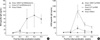
BW-specific IgE levels increased from week 2 through week 5, peaking at week 4 in Group 1 (98.45±64.37 ng/mL) compared with the other groups (Fig. 2A), although the differences between groups were not statistically significant due to the small number of subjects. In a preliminary study we found that sensitizing doses of 10 mg of BW per mouse failed to induce a BW-specific IgE response at any time point between week 1 and 5 after sensitization (data not shown).
HDM-specific IgE responses after intraperitoneal and intranasal HDM sensitization
HDM-specific IgE concentrations increased significantly from week 2 through week 5, peaking at week 3 in Groups 1 and 2 (169.86±55.54 ng/mL, 810.52±233.29 ng/mL, respectively) (Fig. 2B). Interestingly, HDM-specific IgE concentrations in Group 2 were higher than those in Group 1. Repeated administrations of CT and HDM/alum induced significantly higher HDM-specific IgE levels in Group 2. This response was significantly inhibited (79%) by coadministering BW intragastrically in Group 1.
Increased Th2-type cytokine responses
To determine the role of T cells and cytokines in mice allergic to BW and/or HDM, we examined the production of cytokines by splenocytes from these mice. The splenocytes were stimulated in vitro with BW, HDM, Con A, or media. It has been suggested that IL-4 or the balance of IL-4 and IFN-γ plays a key role in regulating the plasticity of both Th1 and Th2 lineage cells, and that this balance is likely to be crucial in regulation of the immune response in vivo (12). After 72 hr in HDM-stimulated culture, IL-4 levels were increased in Groups 1 and 2 (437.16±7.06 pg/mL, 327.79±9.00 pg/mL, respectively) compared with the naive group (37.09±2.23 pg/mL) (Fig. 3A). After 72 hr in HDM-stimulated culture, IFN-γ levels were also increased in Groups 1 and 2 (593.09±60.27 pg/mL, 304.65±17.65 pg/mL, respectively) compared with the naive group (59.45±12.61 pg/mL) (Fig. 3B). However, the differences in IL-4 and IFN-γ levels in HDM-stimulated cultures between Groups 1 and 2 were not statistically significant due to the small number of subjects.
The IL-4/IFN-γ ratio for HDM-stimulated cultures in Group 2 was 1.08, the ratio being slightly higher than those in Groups 1 and 3, implying that CT and HDM/alum-sensitized mice were more skewed toward Th2 response than BW/CT- and HDM/alum-sensitized mice or naive mice. When we normalized IL-4 and IFN-γ levels obtained from splenocytes stimulated in vitro with HDM to those levels obtained from splenocytes stimulated in vitro with media only, the IL-4/IFN-γ ratios for Groups 1, 2 and 3 were 1.1, 1.53, and 0.93, respectively, demonstrating that the IL-4/IFN-γ ratio for CT and HDM/alum-sensitized mice is the highest and that this response was Th2-biased (Table 1). However, IL-5, IL-10, and IL-13 levels in BW-stimulated, HDM-stimulated and unstimulated splenocytes from Groups 1, 2, and 3 showed no significant differences (data not shown).
BW- and HDM-specific IgG1 and IgG2a levels and IgG1/IgG2a ratios
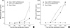
We also measured BW- and HDM-specific IgG1 and IgG2a levels in mice sensitized with BW and/or HDM. BW-specific IgG1 levels increased from week 2 and peaked at week 5 in Group 1 (8,406.5±444.9 ng/mL). BW-specific IgG1 levels showed little increase in Group 2 and in naive mice, as expected (data not shown). HDM-specific IgG1 levels increased from week 2 and peaked at week 5 in Group 1 (40,722.4±9,887.5 ng/mL) and also peaked at week 5 in Group 2 (143,360.5±41,858.5 ng/mL). HDM-specific IgG1 levels showed no increase in the naive group (Fig. 4A).
BW-specific IgG2a levels increased abruptly at week 5 in Group 1 (1,015.2±6.3 ng/mL). BW-specific IgG2a levels showed no increase in Group 2 and in the naive group, as expected (data not shown). HDM-specific IgG2a levels increased from week 3 and peaked at week 5 in Group 1 (110,969.0±2,812.0 ng/mL) and also peaked at week 5 in Group 2 (98,202.2±5,134.8 ng/mL). HDM-specific IgG2a levels showed no increase in the naive group (Fig. 4B).
The ratios of HDM-specific IgG1 to IgG2a for Groups 1 and 2 at week 5 are 0.37 and 1.46, respectively, implying that CT- and HDM/alum-sensitized mice are more Th2-deviated than BW/CT- and HDM/alum-sensitized mice (Table 2).
DISCUSSION
The prevalence of allergic disorders has been rising in recent years, particularly in westernized metropolitan areas. Children with atopic dermatitis or food allergy in early life are more likely to develop asthma or allergic rhinitis later in life, a phenomenon known as "allergic march". In this context, there have been several studies reporting that early sensitization to food allergens in infancy should be regarded as a risk factor for the development of asthma in later years (6, 13-15).
Previously published murine models of food allergy utilized parenteral challenge and therefore did not adequately mimic human food allergy in real life (16). Recently, Li et al. established a murine model of food allergy by utilizing several factors, such as the administration of CT. CT, an enterotoxin from Vibrio cholerae, was found to possess strong T helper cell 2 driving mucosal adjuvant properties, thus mimicking the human IgE responses as well as the clinical symptoms of allergic reactions (9, 17). We applied the well-established factors, including CT, to overcome the strong innate tendency of oral tolerance in mice in our experimental design (17-19). An additional important problem with previously reported murine models is that they focused on either food allergy or inhalant allergy and therefore did not adequately mimic what happens in real life, where sensitizations to foods and inhalants occur almost simultaneously. To our knowledge, this is the first murine model demonstrating the cosensitization of both food and inhalant allergy, generated by intragastric sensitization and intraperitoneal and intranasal sensitization, respectively.
It has been demonstrated that IgE antibodies play an important role in mediating type I hypersensitivity in humans (20, 21). In both food and inhalant allergy it is accepted that food- or HDM-specific IgE binds to high-affinity Fcε RI on mast cells, basophils, macrophages, and dendritic cells, as well as to low-affinity Fcε RII on macrophages, monocytes, lymphocytes, eosinophils, and platelets (22). When food or HDM allergens penetrate mucosal barriers of the gastrointestinal or respiratory tract and contact IgE antibodies bound to mast cells or basophils, histamine and other mediators that induce symptoms of immediate hypersensitivity are released (17).
Von Garnier reported that repeated administrations of low phospholipase A2 doses alone induced a high phospholipase A2-specific IgE level in murine systems (23). This specific response was partially inhibited (36%) by coadministering a low ovalbumin dose and was significantly suppressed (77%) by coimmunization with a high ovalbumin dose (23). These observations thus indicated that coimmunization with an unrelated antigen may exert a significant non-specific bystander effect on the IgE response (23). This non-specific bystander effect is considered to be 'negative' because the unrelated antigens had down-regulatory effects on each other's IgE responses. In contrast to this 'negative' bystander effect, Kullberg and colleagues reported that 'positive' bystander effects occur in murine systems, such that ongoing Th2-dominated immune responses to one antigen enhance Th2 cytokine production in response to other antigens that do not ordinarily induce Th2 cytokines (24, 25). In our experiment where repeated administrations of low doses of HDM alone (Group 2) was introduced, a high HDM-specific IgE level was observed, which was also found to be highly correlated with IL-4/IFN-γ ratio (Group 1 vs. Group 2:1.1 vs. 1.53) and HDM-specific IgG1/IgG2a ratio (Group 1 vs. Group 2:0.37 vs. 1.46). This specific response was significantly inhibited (79%) by coadministering low doses of BW. These observations consequently indicate that cosensitization with unrelated antigens may exert a significant non-specific 'negative' bystander effect on the immune response, regardless of the routes by which the allergens are administered.
This study is different from the previous studies by von Garnier (23) and by Kullberg (24) in that the two antigens used in the present study were administered by methods very similar to the actual routes by which humans become sensitized to the antigens, therefore adequately mimicking human food and inhalant allergies. Thus our study demonstrates that the same pathogenesis might be responsible for the production of BW- and HDM-specific IgE in humans.
Another factor to consider is the endotoxin effect on T cell cytokine production. Endotoxin is known to augment an allergic reaction if administered before or shortly after allergen sensitization and to mitigate allergic reaction if administered at later time points after allergen sensitization (26). Th2 cytokine levels may not have increased as much as we expected because endotoxin might have played a role in the allergic reaction. However, since we have no clear evidence indicating that endotoxin has a direct influence on the cytokine production in this study, this issue should be addressed by future studies. On the other hand, according to our separate experimental data, no additive influence of endotoxin on the proliferative capacity and cytokine productivity of splenocytes in naive or sham control mice was found (27).
Taken together, these findings showed that cosensitization with food (BW) and inhalant (HDM) allergens given by different routes resulted in partial inhibition of the production of HDM-specific IgE. This response is speculated to be mediated by a 'negative' bystander effect, which could be an interesting mechanism to control allergen sensitizations.


 XML Download