

 PDF
PDF ePub
ePub Citation
 Print
Print


INTRODUCTION
Laser in situ keratomileisus (LASIK) is one of the most widely used methods for the correction of refractive errors, because it can lead to rapid recovery of vision with less pain and without occurrence of corneal opacity. However, the creation of the corneal flap can weaken the stroma beneath and there may be an increased risk for keratectasia postoperatively (1, 2).
Even if the factors related in the genesis of keratectasia and the biomechanical changes of cornea after the creation of flap are still being investigated, reduced residual stromal thickness is important as well. There have been several studies insisting that at least a 250 to 300 µm stromal bed should be left to prevent complications (3, 4). However, these prior studies have some limitations; there has been limited consideration for the wide range of thickness of the flap created by one specific microkeratome. In our study, we intended to calculate the estimated rate of the residual stroma of less than 250 µm and investigated if 250 µm is an adequate criterion for preventing postoperative keratectasia statistically.
MATERIALS AND METHODS
We retrospectively investigated the medical records with the proper informed consents of 958 patients (1,916 eyes) who underwent bilateral simultaneous LASIK in Vision Eye Center from April 2000 to October 2003. The finding of keratectasia was defined as marked thinning with the progression of more than 30 µm within 6 months in the area of an ectasia, showing abrupt topographical steepening more than 1.0 diopter (D) and posterior corneal steepening in Orbscan (Orbitek) (5).
The data in this study was analyzed after excluding all other factors, except for residual stromal thickness, that could result in keratectasia. We assumed that keratectasia incidence in our study was the maximum rate that was caused by lack of stromal thickness.
Preoperatively, all the patients were evaluated with slit lamp microscopy, uncorrected (UCVA) and best corrected visual acuity (BCVA), manifest and cycloplegic refraction, keratometry, corneal topography, corneal pachymetry, applanation tonometry and indirect ophthalmoscopy.
All the operations were performed by one surgeon. Proparacaine hydrochloride 0.5% (Alcaine®, Alcon, Fortworth, U.S.A.) was instilled 5 min and just before surgery. We made the expected postoperative total central corneal thickness to be at least more than 400 µm, lest the residual stromal bed should be less than 250 µm in making a 150 µm corneal flap. All the patients had no other ocular or systemic disease that could affect vision after LASIK.
After the creation of superiorly hinged 8.5 mm diameter flap with manual microkeratome (Moria C&B®, 130 head®, Antony, France), 6.0 mm diameter stromal ablation was performed with excimer laser (Schwind Multiscan®, Kleinostheim Germany). Postoperatively, ofloxacin 0.3% (Tarivid®, Santen, Osaka, Japan), fluorometholone 0.1% (ocumetholone®, Samil, Seoul, Korea) were applied 4 times a day for 1 week or more, if necessary. The UCVA, BCVA and the refraction were noted at every visit. The corneal topography and pachymetry were taken at 1 week and 1 month after the operation.
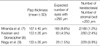
The residual stromal bed thickness was calculated as the remainder from the whole corneal thickness subtracted by the corneal flap thickness. The estimates were based on the ultrasonic pachymetric measurement at 1 week; by this time the flap is stabilized and is not influenced by the desiccation or swelling after surgery (6). The residual stromal bed thickness could be different from the preoperative expected value; this would depend on the real amount of excimer laser ablation and variable flap thickness made by the same microkeratome. The mean flap thickness made by our keratome was 149±24 µm, but is was a small population based data. So we calculated the estimated probabilities for the residual stromal thickness to be below 250 µm using the corneal thickness measured 1 week postoperatively and using the statistical data from the equations below based on the previous studies with the same microkeratome (Table 1) (7-9).
We assumed that the flap thickness constituted a normal distribution (mean µ, standard deviation δ); the total central corneal thickness for the patient n was measured as an, and the flap thickness was X. The mean flap thickness was u, and standard deviation was q for our specific microkeratome based on three different studies (7-9). The probability of residual stromal bed thickness less than 250 µm (Pn) was calculated as follows.
The probabilities were calculated for every patient in the same way (P1, P2, P3 ... Pn). Then the estimated percentage of patients with the residual stromal thickness less than 250 µm in all patients was ΣPn/n. From this equation, we could predict the percentage of the patients whose residual stroma would be less than 250 µm even though we planned, prior to surgery, to make every stromal bed thicker than 250 µm. Then we compared this with the real incidence of keratectasia in our study.
RESULTS
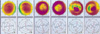
The mean follow-up period was 8 months (range, 1 week to 48 months) and the average age was 31.2±6.1 yr (range, 19 to 53 yr). Preoperatively the mean spherical equivalent was -5.90±1.68 D (range, -1.0 to -12.0 D) and the mean central corneal thickness was 541±31 µm (range, 459 to 640 µm). The mean central corneal thickness at postoperative 1 week was 483±39 µm (range, 371-612 µm). The probability of residual stromal bed thickness less than 250 µm was calculated as 8.8% (168 eyes), using the results from Miranda (u=157, q=40). It was 4.3% (83 eyes) from the results by Kezirian (u=153, q=26) and 1.5% (29 eyes) from Nagy (u=133, q=26). The real incidence of keratectasia in our study was 0.1% (both eyes of 1 patient) in 958 patients (1,916 eyes). The diagnosis of keratectasia was made 2 yr after the operation by topographical measurements (Fig. 1).
The ratios of the real incidence of keratectasia to the percentage of patients whose stromal bed was expected to be less than 250 µm were 1.2% (2/168), 2.4% (2/83) and 6.9% (2/29) respectively according to the data shown above, which was quite low (Table 1). If we plan to make residual stromal bed thickness more than 250 µm using our microkeratome and excimer laser, the iatrogenic keratectasia is less likely to happen.
DISCUSSION
The introduction of laser assisted keratectomy has brought about many associated complications as well as regain of accurate vision (10). Even if the rate of complications has decreased with the development of microkeratome and laser equipment, keratectasia still remains as the most vision threatening side effect.
Different from other types of refractive surgery, some parts of cornea are dissected as a flap before laser ablation in the LASIK procedure. This flap can not act as a barrier against mechanical stress, so iatrogenic keratectasia can easily occur when the remaining stroma is extremely thin, which is particularly a common problem in the surgery of high myopes (11). Since Barraquer insisted that more than 300 µm of stroma should be left to prevent iatrogenic keratectasia, surgical equipment has improved more (12). Therefore, the guideline proposed as the safety margin for residual stroma should be changed accordingly (11, 13, 14).
It is true that the residual stromal bed does not always conform to what we expected before the surgery. In one study of measuring the gap width of 4 different microkeratomes with the same specification of 150 µm, there were significant differences from the manufacturer's specification beyond the standard of tolerance (15). There also can be differences in the degree of laser ablation because the level of energy is not always the same (16). Moreover, the corneal dryness during the surgery also limits the results (17). The preclinical ocular pathology like forme fruste keratoconus and individual differences of corneal tensile strength also make it difficult to determine the safety margin for stromal bed (14, 18). As it is difficult to consider all these factors at a time, and if we assume the thickness of the corneal flap as the only cause of keratectasia, we can estimate the incidence of iatrogenic keratectasia, focusing mainly on the tolerant error of the microkeratome.
Even if the flap is made precisely, it is also difficult to measure the thickness of stromal bed easily during and after LASIK without costly devices. There are several commercial corneal imaging devices to measure residual corneal stromal thickness such as very high frequency ultrasound, optical coherence tomography (OCT), and confocal microscopy (19-21). However, these technologies are too expensive and are not commonly available in many clinical settings. On the other hand, the intraoperative ultrasonic pachymetry is not expensive, however, the accuracy is limited by the fact that the exposed stroma is swollen sometimes or the procedure itself can desiccate the stromal bed (22). As most surgeons do not always measure the residual stromal bed thickness, so we calculated the residual stromal bed indirectly using the postoperative total central corneal thickness and the known variable range of flap thickness. The immediate measurement of corneal thickness after surgery is difficult because there is instability of the flap itself. In addition, the edema of cornea after LASIK can lead to a faulty measurement of corneal thickness, which usually normalizes after the fifth postoperative day (6). The corneal epithelium also has a minute influence on the evaluation of cornea, which becomes thickened between the first and third month postoperatively (23). Therefore, we measured the corneal thickness at one week after surgery, when the edema is mostly absorbed and proliferation of epithelium is not yet prominent.
According to Miranda et al. (7) and Kezirian and Stonecipher (8), the mean flap thickness with the Moria C&B microkeratome was 157 µm and 153 µm, which were not so different. But the expected number of eyes that had a residual stromal bed less than 250 µm was 168 and 83 eyes respectively, which revealed more than a 2 fold difference owing to the different standard deviations. If the standard deviation of flap thickness made by a certain microkeratome is small, we can make a more predictable stromal bed when targeting the same flap thickness. The femtosecond laser maybe a good substitute in this regard (24).
In our study, the real incidence of keratectasia was 0.1%, which was much lower than other reports (4). We think this was because we adhered to the strict protocol to preserve at least 250 µm; in addition, the range of error of our microkeratome and excimer laser might be relatively smaller than those of others.
The minimal adequate stromal bed thickness proposed in our study is at least 250 µm, which was not rigorously applied in the past. If the tolerant errors of microkeratome are considered in planning LASIK, and if we can predict the residual stromal bed more precisely owing to the development of more delicate microkeratome in the future, then the incidence of iatrogenic keratectasia may become lower. But as we could not fully exclude the influence of preclinical ocular pathology like frome fruste keratoconus, and as the mean duration for our study was relatively short, the more studies are necessary.
 XML Download